Showing posts with label pediatric neurologist. Show all posts

Saturday, July 06, 2013

Autism and ADHD Can Occur Simultaneously

HealthDay news imageThis article discusses how autism and ADHD can in many cases occur simultaneously in children. JR

Almost 30 percent of young children with autism also show signs of attention-deficit/hyperactivity disorder (ADHD), a rate that's three times higher than it is in the general population, a new study shows.

"We don't know the cause for ADHD in most cases. We don't know the cause of autism in most cases. It's not surprising that something that's going to affect the brain and cause one developmental outcome may also cause a second developmental outcome," said Dr. Andrew Adesman, chief of developmental and behavioral pediatrics at Steven and Alexandra Cohen Children's Medical Center in Lake Success, N.Y. He was not involved in the study.

Kids in the study who had both problems together also tended to have more difficulty learning and socializing than children who had autism alone.

The researchers noted that the treatment of ADHD may benefit children with autism if they aren't making progress with autism treatment programs, which often require sustained focus on specific skills.

"In a child [with autism] who has great difficulties with attention, or hyperactivity or both, you really have to layer in another level of intervention strategies for them," said study author Rebecca Landa, director of the Center for Autism and Related Disorders at the Kennedy Krieger Institute in Baltimore.

For the study, which is published in the June 5 online issue of the journal Autism, researchers as
ked parents of kids enrolled in a community-based study of child development about symptoms of attention and hyperactivity -- whether or not children could wait their turn, interrupted others who were speaking, fiddled with things during meals or could not slow down, for example. All the children in the study were between the ages of 4 and 8.

Out of 62 children diagnosed with autism, 18 (29 percent) also showed signs of ADHD.

A previous study of slightly older children found that 31 percent of children had the two disorders together.
"It's not surprising," said Dr. Patty Manning-Courtney, director of the Kelly O'Leary Center for Autism Spectrum Disorders at Cincinnati Children's Hospital Medical Center.

"What's good about this study is that they went to the trouble to look at who met diagnostic criteria and what was different about those children," said Manning-Courtney, who was not involved in the research.

All the children who had both problems together were boys. Boys have higher rates of autism and ADHD than girls, research shows.

One limitation of the study was that researchers had to rely on questionnaires that are meant to spot ADHD in typical children. There really aren't good tests for attention and hyperactivity developed for kids with autism, and their problems may look different than those seen in typical school-aged children.

Children who are higher functioning on the autism spectrum can have fairly obvious problems with attention.

"It's not that they have a deficit of attention. It's that they can't allocate their attention, or shift their attention to what it needs to be on," Courtney-Manning said. "I talk to parents of kids with autism about attention regulation more than attention deficit."

In children who are more severely autistic, ADHD can be harder to spot.

"It's hard to tell if their activity level is different because they're delayed or they're more severely autistic or if it's ADHD," Courtney-Manning said.

But if parents and teachers are noticing that attention or activity problems are interfering with a child's ability to make progress, that's the time to seek help, she noted.

First-line treatments for attention problems in kids with autism involve behavioral interventions that aim to teach kids to better control their focus.

If the behaviors don't get better with help, Landa said doctors will then move on to medication.

"If your child is having those kinds of problems, it's worth mentioning to the child's doctors and also touching base with the child's teachers," Landa said.

Read more here

Tuesday, August 28, 2012

The Best Ways To Integrate Special Needs Students


I found this report on NPR. I hear parents talk about special education issues every day. Accommodations and modifications are a difficult process for all. Children with seizures, autism, cerebral palsy, ADHD and other neurodevelopmental problems are entitled to education. Parents expect a full range of services. In general, schools have motivated and highly professional personnel and accurate services. At the same time, schools have limited budgets for personnel, occupational, physical and speech therapy.  Its a difficult situation.  A physician can help. JR


The Best Ways To Integrate Special Needs Students


May 2, 2012
Budget cuts in many school districts have some parents and teachers questioning whether they have the resources to support their students. NPR education correspondent Claudio Sanchez and Thomas Hehir of Harvard University talk about how to integrate special needs students into mainstream classrooms.
JENNIFER LUDDEN, HOST:
This is TALK OF THE NATION. I'm Jennifer Ludden, in Washington. A troubling new video has reinvigorated the debate over special needs education. A father was told his special needs child was unruly, so he sent him to school wearing a wire. On the recording, his son is harassed and mocked by a teacher and aide in a special needs classroom.
The video is only the latest example of a widespread sense of frustration about special education. What to do? Advocates often want special needs students to get their own classes, but school districts say that's expensive, and certainly in a time of budget cuts, mainstreaming is on the rise.
If you're the parent or teacher of a special needs student, what works? Our number is 800-989-8255. Our email address is talk@npr.org. And you can join the conversation at our website. Go to npr.org and click on TALK OF THE NATION. Later in the program, why some schools are forcing kids off their bikes and into cars.
But first, best practices for mainstreaming special needs students. Joining me now is NPR's education correspondent Claudio Sanchez here in Studio 3A. Hi, Claudio. Welcome.
CLAUDIO SANCHEZ, BYLINE: Good to be here.
LUDDEN: So this is a trend that's been growing in public schools, but mainstreaming may not be a term that everyone's familiar with. Can you just tell us what it means?
SANCHEZ: Mainstreaming means that a child's instruction takes place in a classroom with non-disabled kids, regular kids, a decision based often on the special needs of that child. So, yes, there's an attempt to have this kid feel inclusive, be inclusive, but often it has to be tailor-made. That's where something very crucial here has to happen, and that is that an individual education plan be designed for this child, which calls for special accommodations, certainly special attention if necessary.
And this is all, of course, in the law, the 1975 law that actually was originally called the Education for All Handicapped Children's Act. It later became the Individual Disability Education Act, and that was, for the most part, kind of reauthorized in 1990.
LUDDEN: So, for decades, there's been this effort - so there was a sense beforehand that special needs kids were kind of shunted away and kept out of sight. Parents wanted them in the mainstream. And yet, as I understand it, there have been a lot of bad experiences in these mainstream classes.
SANCHEZ: And remember, they weren't just shunted away. They were literally kept out of school. I mean, for many, many years, these kids were warehoused somewhere else. Schools didn't deal with them. You know, it took several legal cases and challenges to that, most - especially in 1972 in Pennsylvania, which literally led to the creation of the law.
But you're right. The problem has been money, in many ways. You know, there's been an acceptance that these kids can learn, should be mainstreamed, but the money issue is huge. The federal government, when it authorized this law, more recently said we're going to contribute 40 percent - this is Congress talking - 40 percent of the funding for special education.
To this day, it's never been more than 18 percent. So that means that local and state education folks have to come up with the money somewhere, and we're talking about tens of thousands of dollars for every child, every year. So it's a very - it's a very difficult problem for schools because they don't have the money.
LUDDEN: And I guess more so now with - we've seen so many cuts at local school districts.
SANCHEZ: Exactly. And certainly in this time of austere and very limited budgets, I mean, where do you go? The federal government is not coming up with more money, believe me. States are obviously cutting left and right. So, I mean, often, it comes down to litigation on the part of parents. And if you have a good attorney, sometimes they get money out of the district to pay for these services or to put the child in a private program.
LUDDEN: So this terrible, painful video has surfaced of this child's experience. But I take it that's not a surprise to people who look at this field, that there's been a series of studies recently that show this these kinds of instances.
SANCHEZ: Yes, although I would say that the - you know, it's difficult to really document, certainly, every instance of abuse, but they are pretty common. You know, there are cases - there was on in Georgia, a 13-year-old boy committed suicide after being sent to an eight-by-eight, concrete-block time-out room in Gainesville, Georgia, at the public school there.
For students, this was a place that they put students in for behavioral problems. Then there was - you know, there are these famous screaming rooms that some schools have where teachers put kids when they're acting out, when they're out of control. And, you know, there was that famous case, I forget where, it may have been Kentucky, where a child was - who was misbehaving, a special ed kid, had been found stuffed in a duffel bag.
I mean, you know, you hear about these things, and you say this can't be. This has to be the exception to the rule. But you'd be surprised how often - I mean, some of these things aren't even reported, but it happens.

Saturday, May 19, 2012

Just What's Inside Those Breasts?


breasts.jpgJust What's Inside Those Breasts?

May 16, 2012

When writer Florence Williams was nursing her
second child, she read a research study about
toxins found in human breast milk. She decided
to test her own breast milk and shipped a
sample to a lab in Germany. What came back surprised her.

Trace amounts of pesticides, dioxin and a jet
fuel ingredient — as well as high to average
levels of flame retardants — were all found in
her breast milk. How could something like this
happen?

"It turns out that our breasts are almost like
sponges, the way they can soak up some of these chemicals, especially the ones that are fat­
loving — the ones [that] tend to accumulate in fat tissue," Williams tells Fresh Air's Terry Gross.
"Unfortunately, the breast is also masterful at converting these molecules into food in the way of
breast milk."

Learning that breasts soak up lots of chemicals made Williams wonder just what else was going on
with breasts. A lot, as it turns out. In her new book, Breasts: A Natural and Unnatural History,
Williams offers her take on — among other things — why breasts are getting bigger and
developing earlier, why tumors seem to gravitate toward the breast, and how toxins from the
environment may be affecting hormones and breast development.

She says many of those toxins, including the flame retardants found in her breast milk, may come
from ordinary household items like couches and electronics, which often contain flame retardants.
Some animal studies have shown that certain types of flame retardants interact with hormone...

Listen here

Long-term Treatment Outcomes of Children&Adolescents who have Cerebral Palsy with Secondary Osteoporosis.


Editor's Note:  I start screening my most severe patients and teenagers for osteoporosis when treating cerebral palsy. JR

Curr Med Res Opin. 2012 May;28(5):737-47. Epub 2012 Apr 18.

Long-term outcomes of children and adolescents who had cerebral palsy with secondary osteoporosis.

Source

Kitasato University School of Medicine , Sagamihara , Japan.

Abstract

Abstract Objective: To investigate the long-term efficacy and index of treatment with vitamin D alone or with a bisphosphonate in children and adolescents who have cerebral palsy (CP) with secondary osteoporosis. Research design and methods: Thirty patients diagnosed with CP and secondary osteoporosis were analyzed for 5 years, and the efficacy of treatment was compared. Treatment was divided into three groups: The monotherapy group, consisting of patients taking only alfacalcidol (0.03 µg/kg/day); the polytherapy group, consisting of those taking alfacalcidol and risedronate (0.05 mg/kg/day); and the control group, consisting of patients who discontinued taking their medications for reasons unrelated to these therapies. Bone mineral density (BMD), bone-specific alkaline phosphate (BAP), and N-telopeptides of type I collagen (NTX/Cr) were measured on each patient just before and at discontinuation of treatment, after 6 months, and again at 1 and 3 years, respectively. The changes in BMD (ΔBMD), BAP (ΔBAP), and NTX/Cr (ΔNTX/Cr) were evaluated at these intervals, because the normal value of each parameter varies over time during childhood. Results: ΔBMD significantly increased in the polytherapy group at ≥1 year (p = 0.006), and the difference in BMD between the polytherapy and the control groups at ≥1 year was also significant (p = 0.005). ΔBAP was increased in the monotherapy and polytherapy groups at ≥1 year (p = 0.021 and p = 0.033). ΔNTX/Cr decreased in the polytherapy group at ≥1 year, which was consistent with the polytherapy group of the period from 1 month to 1 year (p = 0.033). The relation between ΔBMD to ΔBAP was a positive correlation in the second period in the monotherapy group (r = 0.46). And the relations between ΔBMD to ΔNTX/Cr were not recognized negative correlations in the monotherapy and polytherapy groups. Thus, ΔBMD reflected ossification of secondary osteoporosis in patients with CP, and ΔBAP and ΔNTX/Cr was significantly related to the increase and decrease of ΔBMD. There were no effects of other factors except sexual maturity. Limitations of this study include that each index of examination was the evaluation according to rate of change. Therefore, the results of this study were limited to longitudinal evaluations. Conclusion: Evaluation according to ΔBMD and both methods of monotherapy and polytherapy were useful for CP patient taking antiepileptic drugs (AEDs) and regardless of sex. Especially, polytherapy for longer than 1 year led to improvement in BMD in children who had CP with secondary osteoporosis. BAP and NTX/Cr were useful for the index of the progression osteoporosis with or without these therapies.


Read More: http://informahealthcare.com/doi/abs/10.1185/03007995.2011.645562

Boy with Cerebral Palsy Walks to his Marine Dad Returning from Deployment


A boy with cerebral palsy was told he'd never walk, but did just that when he saw his returning Marine dad.


http://video.insider.foxnews.com/thumbnails/694940094001/2012/05/10/640/360/694940094001_1631328903001_vs-1631314523001.jpg

link here

Thursday, March 29, 2012

Did you know that one quarter of babies born by normal vaginal birth will have an asymptomatic brain hemorrhage?

Intracranial Hemorrhage in Asymptomatic Neonates: Prevalence on MR Images and Relationship to Obstetric and Neonatal Risk Factors1

Newborn on yellow blanket being attended to by a nurse


Christopher B. Looney, BS, J. Keith Smith, MD, PhD, Lisa H. Merck, MD, MPH, Honor M. Wolfe, MD, Nancy C. Chescheir, MD, Robert M. Hamer, PhD and John H. Gilmore, MD

+ Author Affiliations

1From the Department of Psychiatry, CB No. 7160, 7025A Neurosciences Hospital, University of North Carolina School of Medicine, Chapel Hill, NC 27599-7160. From the 2005 RSNA Annual Meeting. Received January 23, 2006; revision requested March 23; revision received June 7; accepted June 21; final version accepted August 21. J.H.G. supported by National Institute of Mental Health grant 1 P50 MH064065. C.B.L. supported by a Distinguished Medical Scholarship from UNC School of Medicine.

Address correspondence to

J.H.G. (e-mail: jgilmore@med.unc.edu).

Next SectionAbstract

Purpose: To retrospectively evaluate the prevalence of neonatal intracranial hemorrhage (ICH) and its relationship to obstetric and neonatal risk factors.

Materials and Methods: Pregnant women were recruited for a prospective study of neonatal brain development; the study was approved by the institutional review board and complied with HIPAA regulations. After informed consent was obtained from a parent, neonates were imaged with 3.0-T magnetic resonance (MR) imaging without sedation. The images were reviewed by a neuroradiologist with 12 years of experience for the presence of ICH. Medical records were prospectively and retrospectively reviewed for selected risk factors, which included method of delivery, duration of labor, and evidence of maternal or neonatal birth trauma. Risk factors were assessed for relationship to ICH by using Fisher exact test statistics.

Results: Ninety-seven neonates (mean age at MR imaging, 20.8 days ± 6.9 [standard deviation]) underwent MR imaging between the ages of 1 and 5 weeks. Eighty-eight (44 male and 44 female) neonates (65 with vaginal delivery and 23 with cesarean delivery) completed the MR imaging evaluation. Seventeen neonates with ICHs (16 subdural, two subarachnoid, and six parenchymal hemorrhages) were identified. Seven infants had two or more types of hemorrhages. All neonates with ICH were delivered vaginally, with a prevalence of 26% in vaginal births. ICH was significantly associated with vaginal birth (P < .005) but not with prolonged duration of labor or with traumatic or assisted vaginal birth.

Conclusion: Asymptomatic ICH following vaginal birth in full-term neonates appears to be common, with a prevalence of 26% in this study.


link

Monday, February 27, 2012

Use of cooling blanket spurs new infant brain research into optimal care


Pediatric neurologists and neonatologists in Vanderbilt’sNeonatal Intensive Care Unit(NICU) are conducting research that may better define what happens in the brains of newborns who have suffered from oxygen deprivation, and what brain cooling therapy can achieve.
Use of a “cooling cap” within six hours is now the standard of care when an otherwise healthy, full-term infant experiences a serious lack of oxygen, called anoxic brain injury. The therapy uses cool water to bring the temperature of the brain down about five degrees. Vanderbilt took part in the initial studies of the cooling cap, which was found to reduce serious long-term brain damage and death by about one-third.
Now a switch has been made to a cooling blanket instead of a cap. The blanket is as effective in its brain-protecting capacity, but it offers researchers an opportunity to better assess what is happening in the brain as it is cooled.

Easier to monitor babies’ brains

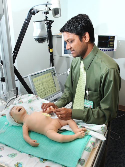
Pediatric neurology chief resident Siddharth Jain said by using a cooling blanket, the scalp is free for him to apply a full, nine-lead electro encephalogram (EEG) monitor along with a newer device called a near-infrared spectroscopy (NIRS) monitor to find out what is going on beneath a baby’s scalp.
The EEG can detect seizures, while NIRS measures how the brain uses oxygen. The information collected so far has been surprising.
“We already know the EEG detects seizures that cannot be seen in these babies. Up to 80 percent of seizures in babies with anoxic brain injury have no overt clinical signs. Other studies have shown that these babies can have between 100 and 120 seizures during the first 72 hours of life,” Jain said.
“These seizures are different than those in epilepsy. They are very aggressive in the first 48 hours and difficult to control. We don’t know for sure, but the consensus is the seizures themselves cause further damage.”
Jain is working with Barbara Engelhardt, associate professor of neonatology, to closely examine what is happening in the first 72 hours after an anoxic injury — the critical window of opportunity in which the cooling seems to have its maximum brain-preserving effect.
For this observational study, William Walsh, neonatologist and director of nurseries, switched the NICU to the cooling blanket to allow the researchers to enroll the one or two babies brought to Vanderbilt every month with anoxic brain injury.

Speeding up evaluation and treatment

The first goal is to use EEG and NIRS to more quickly and accurately predict the severity of the anoxic injury. Currently, an MRI is used to give doctors a peek at structural damage in the brain, once the initial injury has stabilized, to assess long-term changes. But the predictive value of long term damage is not very great.
Engelhardt says EEG and NIRS provide information from day one about seizure activity as well how the brain is using oxygen.
These two pieces of information can describe changes after the initial injury, and may help provide a better measure of the effects on long-term outcomes.

Enables researchers to test new therapies

A second goal is to use the monitors to test new therapies. The hope is the monitors could more quickly assess the effectiveness of therapies is controlling seizures, or better balancing nutrient consumption after injury in the hope of further reducing the risk of permanent brain damage.
“When a brain cell is injured, it can go one of two ways: it can die or recover,” Walsh said. “The goal is to limit damage to as small an area as possible.
“The cooling cap was a general way to slow down the potential for further damage, but in the 15 years we have been using it, we have learned a lot about what is happening biochemically in anoxic injury and how we can further impact that damage.”

Saturday, February 11, 2012

Why Brain Injuries Are More Common In Preemies


Why Brain Injuries Are More Common In Preemies


The most common cause of brain injury in premature infants is a lack of oxygen in the days and weeks after birth, researchers say.
Scientists say they are beginning to understand why brain injuries are so common in very premature infants — and they are coming up with strategies to prevent or repair these injuries.
The advances could eventually help reduce the number of premature babies who develop cerebral palsy, epilepsy or behavioral disorders such as ADHD, researchers told the Society for Neuroscience meeting in Washington, D.C., this week.
Each year more than 60,000 babies are born weighing less than 3.3 pounds. And because of advances in neonatal medicine over the past several decades, most of those babies will survive. But researchers have had less success finding ways to prevent brain damage in these infants.
"That means that overall rates of cerebral palsy and other neurodevelopmental disabilities are on the rise," says David Rowitch, chief of neonatology at the University of California, San Francisco.
The most common cause of brain injury in premature infants is a lack of oxygen in the days and weeks after birth, Rowitch says. The lack of oxygen damages white matter, which provides the "communication highways" that carry messages around the brain and to distant parts of the body, he says.
 
And the babies at greatest risk of this sort of brain damage are those born after as little as six months of gestation, Rowitch says.
"Such a baby would weigh about a pound and would fit into the palm of your hand," he says. "As you can imagine, they're very fragile and vulnerable to stresses."
Those stresses often include periods when an infant's immature lungs are not delivering enough oxygen to the brain, even with help from a mechanical breathing device.
This lack of oxygen appears to damage the most common type of white matter, myelin, which acts like an insulator around the nerve fibers that carry messages in the brain and nervous system. Without enough myelin, short circuits can prevent these messages from getting through, Rowitch says.
He initially found evidence of white matter damage by studying brains from premature infants who died. But since then, he's been able to assess premature infants using a special incubator designed to fit in an MRI scanner.
"We've been able to now take over 250 babies who are very preterm to the MRI scanner safely to show that this is a feasible way to detect white matter injury early on," he says.
Now the question is how to prevent or repair that sort of injury.
Some studies show that it's important to act right away, says Vittorio Gallo from Children's National Medical Center in Washington, D.C.
"There is a very critical developmental time window right after birth," Gallo says. "If development is disturbed during this critical time window then the brain doesn't catch up."
Gallo is part of a team of scientists who have shown that it is possible to intervene — at least in mice. One approach involves giving the mice a drug that speeds up production of myelin, he says.
"We do this intervention right after the injury," he says. "And we found that by targeting specific targets we can recover and regenerate at least part of these cells right after the injury, during that critical developmental time window."
Any drug for people is still years off, Gallo says
But other scientists at the meeting say there are promising treatments available now. These include everything from the magnetic stimulation of certain areas of the brain to temporarily lowering the body temperature of premature infants to protect brain tissue.


Full story here

Thursday, December 08, 2011

Study Finds Headaches after Traumatic Brain Injury Highest in Adolescents and Girls


More than half a million children in the U.S. sustain a traumatic brain injury (TBI) every year. Adults who suffer TBI often report headaches afterward, but little is known about how often children suffer headaches after similar injuries. In a significant new study, “Headache After Pediatric Traumatic Brain Injury: A Cohort Study,” researchers analyzed the prevalence of headaches three and 12 months after mild, moderate or severe TBI in children ages 5 to 17, and discovered the risk of headache was higher in adolescents (ages 13 to 17) and in girls. The study was led by Heidi Blume, MD, MPH, from Seattle Children’s Research Institute and principal investigator Fred Rivara, MD, MPH, of Harborview Injury Prevention and Research Center, University of Washington, and is published online in Pediatrics.

The study indicates that headache can be a significant problem for some children (ages five to 12 years) after TBI. Three months after a mild TBI, 43 percent of children reported headaches, compared to 37 percent of children who had a moderate to severe TBI, and 26 percent of children in the control group (patients with arm fractures).

Study authors conclude that the response to and recovery from TBI is different for children, adolescents and adults, and that males and females are likely to have different symptoms and recovery. The risk of headache was higher in adolescents and in girls, mirroring a pattern seen in other headache disorders such as migraine. Because of the high number of children suffering TBI every year, the study findings indicate many children and adolescents are suffering from TBI-associated headaches every year.

“Little research has focused on chronic headache post-TBI in children,” said Dr. Blume, who is also a University of Washington assistant professor of neurology. “Our findings indicate that many children and adolescents suffer from TBI-associated headaches yearly. In addition, the prevalence of headache following mild TBI appears to follow a pattern we see in primary headache disorders such as a migraine. With future research, we can begin to examine whether there are similarities in the cause of migraine and post-traumatic headache, and if migraine therapies will work for post-traumatic headaches.”

Researchers were not able to detect significant differences in the percentage of children with headache after TBI one year after injury, compared to children with arm fractures. The study concluded that adolescents and girls appear to be at the highest risk for headache after mild TBI, and that the course of recovery from TBI is likely affected by age at injury, injury severity and gender.

What parents need to know is that some children with TBI may have headaches for several weeks or months after TBI, but that most recover with time,” said Dr. Blume. “And significantly, girls and teenagers appear to be at particular risk for headaches after mild TBI. Parents should be aware of what to expect after mild TBI, which may come from a sports-related injury.”

If you suspect your child has had a concussion, see your medical care provider before allowing them to go back to vigorous physical activity or playing sports. Seek emergency care after a head injury if the child has repeated vomiting or severe new headache, is confused, off balance, or has new weakness, numbness or trouble speaking.

To manage headaches in children and adolescents, Dr. Blume recommends these SMART tips:

  • Sleep – get regular and sufficient sleep (Eight to nine hours/night)
  • Meals – eat regular and healthy meals, including breakfast and drink plenty of fluids to stay well hydrated
  • Activity – get appropriate activity, rest immediately after TBI, but if symptoms persist for several weeks your care provider may recommend supervised regular low impact exercise such as walking that does not exacerbate symptoms
  • Relaxation – manage stress and find ways to relax
  • Trigger avoidance – avoid things that make headaches worse (loud noise, bright lights, stress, skipping meals, sleep deprivation)

Read more: http://www.biosciencetechnology.com/News/2011/12/Study-Finds-Headaches-after-Traumatic-Brain-Injury-Highest-in-Adolescents-and-Girls/

Sunday, August 07, 2011

Concussions Are Now Priority One on School Football Fields


Concussions have been so roundly ignored on the football field that a law is now in effect to force their treatment. Joshua Rotenberg M.D. of Texas Medical and Sleep Specialists says there are nearly four million concussions a year in the country. He says so often concussions are overlooked, that there needed to be a law.

"Every school district that has interscholastic sports, they have to remove a child from play, or practice, if they suspect that they have had a concussion."

He says the demands of football practice and the priority that Texas places on sports often supercedes treatment on the field.

"People are so motivated to play the game, that not even an alteration in their brain function will stop them."

Rotenberg says a concussion can remain symptomless ... hours after the injury. Symptoms include headache, nausea and being forgetful. This means kids will continue to play, making things worse.

"It gets worse with exercise so after sitting on the sidelines for a few minutes, you might feel fine but then a kid gets back in the game and they end up messing up a play that they normally would have done just fine."

He says such an injury, left untreated, can lead to permanent brain damage.


http://ccsanantonio.com/cc-common/news/sections/newsarticle.html?feed=119078&article=8909918

Sunday, July 31, 2011

New Study Looks At Football Helmets And Concussions

New Study Looks At Football Helmets And Concussions

Check out the new rankings at www.sbes.vt.edu/nid.php

By TIM LAMMERS

FOX CT

7:21 PM EDT, July 30, 2011

If you have a child playing high school or college football, then this story is for you. A new study looks at helmets and which ones might be the safest for your child. But not everyone agrees on the results.

Concussions have always happened in football. But only recently have we started to discover just how dangerous they really are.

"Concussions are the number one, perhaps the number one interest, and therefore the number one concern," says Dr. Carl Nissen, Elite Sports Medicine.

So it was only a matter of time before football helmets came under closer scrutiny as well.

"There is, probably, some promise that the newer helmets can reduce the rate of concussions," says Dr. Nissen.

The current standard for NFL and a lot of college, and even high school, teams comes from the National Operating Committee on Standards for Athletic Equipment (NOCSEA). So as long as you've got their seal of approval, you're good to go.

"If a helmet has been approved by NOCSAE, it is gonna meet the minimal standards, so it is gonna be a protective piece of equipment," says Dr. Nissen.

Well, a brand new study has reached some drastically different conclusions.

"The study that we're all talking about as we go into this fall's football season is the STAR criteria for helmet safety," Dr. Nissen says. "A sliding scale of one through five stars as to whether a helmet is good or bad, safe or not."

Like the five-star rated Riddell Revolution Speed. That and all the four-starr rated helmets are highly recommended.

"Their techniques are very well established," says Dr. Nissen. "Their lab does this very well."

But two helmets were not recommended for use. One of them, the Riddell VSR4, is still commonly found in the the NFL. It also has NOCSAE's approval. And NOCSAE isn't changing its stance because Virginia Tech has yet to finish real-world testing to prove that concussion rates match up with the star ratings.


http://www.courant.com/health/connecticut/ctn-new-study-looks-at-football-helmets-and-concussions-20110730,0,3685152.story

Football players - Check out Helmet Ratings

Adult Football Helmet Ratings - May 2011

A total of 10 adult football helmet models were evaluated using the STAR evaluation system for May 2011 release. All 10 are publicly available at the time of publication. Helmets with lower STAR values provide a reduction in concussion risk compared to helmets with higher STAR values. Based on this, the best overall rating of ‘5 Stars’ has the lowest STAR value. Group rankings are differentiated by statistical significance.

http://www.sbes.vt.edu/nid.php

How expensive is autism?

Here is a very sobering post by my colleague and friend, Pat Harkins MD. She walks the walk...


How expensive is autism?

Posted by Patricia Harkins in Uncategorized


The short answer is “very”.

The long answer is, well, long

Note – I use ASD to mean “autism spectrum disorder”.

Let’s start with therapy. I estimate the average ASD child needs Speech/Language therapy for about 5 years and Occupational Therapy for about 3 years. That is 16 “therapy years”. This is about $300K to the family or insurance. The cost to public schools is about $4K per year for 13 years. Cost in babysitters because they can’t go to a regular daycare is about $5K to 26K per year needed, perhaps for average of 10 years...

In our family’s case, my husband stopped being employed when our son was 3 because of his needs

....read more at

http://autismwhisperer.net/2011/07/30/how-expensive-is-autism/

Thursday, July 21, 2011

'Laughing gas' sedation calms pediatric hospital patients

'Laughing gas' sedation calms pediatric hospital patients

I am at my cerebral palsy/ "Botox" clinic today in san Antonio. Its amazing to see how cal kids are with sedation. Laughing gas makes this quick and easy.

SAN ANTONIO -- Doctors at a San Antonio hospital are using a new tool to make their smallest patients more comfortable. It’s a sedation method dentists have used for years.
This is a less invasive way to sedate children for procedures. All it requires is a mask and about three minutes.

Keeping the smallest of patients calm and tranquil during procedures is no small task. Many diagnostic or therapeutic interventions require IV sedation where children are unconscious. For some patients, though, there’s another less invasive alternative.

Gas. It’s nitrous oxide, the same stuff you’d call “laughing gas” at the dentist.

At North Central Baptist Hospital in Stone Oak, the pediatric sedation unit has been using this approach of the last two months with great success.

“They’re awake the whole time,” explained Dr. Daniel Sedillo, a pediatric critical care specialist. “You can talk to them and tell them to take a deep breath or to relax and once you take the gas off within two or three minutes they’re back to their playful self as they were before.”

The gas can be kept up for about half an hour and allows doctors to do everything from injections to biopsies, from spinal tapes to MRI scans.

The children have less angst and fear and the doctors have a cooperative patient to work on.

“The physicians who were doing the procedure were very happy that they were able to do the procedure comfortably without the child moving a whole lot and not having to see tears or screaming or anything like that,” Sedillo remarked.

Using nitrous oxide as a sedative means parents don’t have to worry as much about what their children eat before the procedure. The recovery to full consciousness is faster.


http://www.kens5.com/home/Laughing-gas-sedation-calms-pediatric-hospital-patients-103462504.html

Monday, July 18, 2011

Laser Cell Ablation for Epilepsy - Read Keagan's Story


Read Keagan 's Story


Laser Cell Ablation for Epilepsy

Texas Children's Hospital is the first hospital in the world to use real-time MRI-guided thermal imaging and laser technology to destroy lesions in the brain that cause epilepsy and uncontrollable seizures.


According to hospital experts, this new surgical approach offers a safer and significantly less invasive alternative to craniotomy, currently the most commonly used cranial surgical treatment for epilepsy. For high-risk patients with deep brain lesions, this new technique can be particularly life-changing because the MRI-guided laser probe utilizes a much smaller pathway through the brain to reach a deep lesion. This reduces the risk of patient complications related to contact with surrounding brain tissue. In addition, the MRI-guided laser probe is inserted through a hole in the skull that is only 3.2 mm (about the diameter of a pen) versus the removal of a larger area of skull bone for a craniotomy. Because it is a less invasive procedure, patient recovery time is much shorter.


Keagan's StoryA recent example of the effectiveness of this new surgery is nine-year-old Texas Children’s Hospital patient Keagan Dysart, of Converse, Texas, who suffered from two types of epileptic seizures when he was diagnosed with a hypothalamic hamartoma in his brain. The gelastic seizure caused him to giggle and laugh uncontrollably two or three times an hour. Keagan would also periodically experience a tonic seizure, with generalized body stiffening and loss of awareness that caused him to fall asleep for sometimes up to an hour afterward.
Keagan’s case was particularly high risk because his lesion was located in the hypothalamus, near the brain stem. In this highly sensitive region, there are a myriad of potential, serious complications from surgery including loss of sight, damage to the pituitary gland, stroke from artery damage or development of diabetes insipidus (DI), a potentially fatal condition where the kidneys are unable to conserve water because of disruption to the area of the brain that releases the body’s anti-diuretic hormone.
The location, size and complexity of Keagan’s brain lesion made him an ideal candidate for the new surgical procedure, which was successfully performed in March without any surgical complications. He is now seizure-free.
View a presentation describing the breakthrough epilepsy procedure.


Texas Children’s Hospital pioneers use of MRI-guided laser surgery for revolutionary new epilepsy treatment

Read about the story of one of my patients!!! JR


Texas Children’s Hospital pioneers use of MRI-guided laser surgery for revolutionary new epilepsy treatment


New procedure significantly reduces risk of patient complications and recovery time


HOUSTON – (July 18, 2011) – Texas Children’s Hospital is the first hospital in the world to use real-time MRI-guided thermal imaging and laser technology to destroy lesions in the brain that cause epilepsy and uncontrollable seizures.

News media contact
Christy Brunton832-824-2645clbrunto@texaschildrens.org
According to hospital experts, this new surgical approach offers a safer and significantly less invasive alternative to craniotomy, currently the most commonly used cranial surgical treatment for epilepsy. For high-risk patients with deep brain lesions, this new technique can be particularly life-changing because the MRI-guided laser probe utilizes a much smaller pathway through the brain to reach a deep lesion. This reduces the risk of patient complications related to contact with surrounding brain tissue. In addition, the MRI-guided laser probe is inserted through a hole in the skull that is only 3.2 mm (about the diameter of a pen) versus the removal of a larger area of skull bone for a craniotomy. Because it is a less invasive procedure, patient recovery time is much shorter.
More than three million people in the U.S. are affected by epilepsy, including about 300,000 children under the age of 14. Uncontrollable seizures can affect a child’s memory, concentration, motor skills, school performance and quality of life. Drug therapies control seizures in about 60 percent of those with epilepsy. Invasive craniotomy, nerve stimulation and special diets are treatment options for patients who do not respond to medication.

Studies associate head injuries with dementia in later life.

Studies associate head injuries with dementia in later life.

The Wall Street Journal (7/18, A3, Wang, Subscription Publication) reports that, according to two studies to be presented today at the Alzheimer's Association International Conference on Alzheimer's Disease in Paris, concussions and other head injuries may be associated with dementia in later life and may also increase vulnerability for Alzheimer's symptoms. In the first study, researchers who analyzed data on 280,000 veterans of the military older than age 54 found that those who had a diagnosis involving traumatic brain injury faced triple the risk for developing dementia. In a second study, which involved about 500 former professional football players with the NFL who were showing symptoms of mild cognitive impairment, researchers found that the players had similar cognitive function scores as elderly people with MCI, even though the players were years younger.

An AP (7/18, Marchione) story run by at least 146 publications reports, "A large study in older veterans raises fresh concern about mild brain injuries that hundreds of thousands of American troops have suffered from explosions in recent wars. Even concussions seem to raise the risk of developing Alzheimer's disease or other dementia later in life, researchers found." The research, funded by the US Department of Defense and the National Institutes of Health, was led by Dr. Kristine Yaffe, director of the Memory Disorders Clinic at the Veterans Affairs hospital in San Francisco. Researchers working on the study "reviewed medical records on 281,540 veterans who got care from Veterans Health Administration hospitals from 1997 to 2000 and had at least one follow-up visit from 2001-2007."

According to the Wall Street Journal (7/18, Wang, Subscription Publication), Yaffe's says her findings raise concerns about the possible long-term impact of veterans' traumatic brain injuries. The Journal also took note of a separate study which found that professional football players may also be at increased risk of cognitive problems later in life. USA Today (7/18, Marcus) notes, "Yaffe says more research is needed to explore whether early rehabilitation can help reduce the risk for dementia." She added, "If you know you've had a head injury and you are approaching older age, one has to be carefully monitored and screened for cognitive dementia."

Bloomberg News (7/18, Torsoli) notes that Yaffe's research findings will be "presented at the Alzheimer's Association International Conference in Paris" on Monday, where they will "highlight another potential hazard of war. Traumatic brain injury is a 'signature wound' of the conflicts in Iraq and Afghanistan, accounting for 22 percent of casualties overall and 59 percent of blast-related injuries, said" Yaffe.

http://yourlife.usatoday.com/health/medical/alzheimers/story/2011/07/Traumatic-brain-injury-doubles-risk-of-Alzheimers/49472436/1?dlvrit=205764

Sunday, July 17, 2011

High annual concussion count calls for safety - New Texas Laws

Natasha's Law to make debut this season

High annual concussion count calls for safety

By JASON McDANIEL
For the Chronicle

July 16, 2011, 11:29PM


Cartoonist Steve Moore has devoted plenty of ink to lampooning everything associated with athletic training in his nationally syndicated comic In the Bleachers.

But even he admitted questioning whether to make light of concussions during his keynote address this week at the Southwest Athletic Trainers Association's 57th meeting at the Westin Galleria.

Of course, he still does it.

The cartoon he brought featured a machine that sucks concussions out of an athlete's head, and it's funny. But his hesitation underscores the seriousness of the issue. That goes double for high school athletes, whose brains still are developing.

"If a youngster sprains his ankle, the parents, coaches and the youngsters themselves can see the swelling, they can see the limping, the fact that they're on crutches and so forth, so they can follow the injury and understand the concept," said Bucky Taylor, an athletic trainer for Mesquite ISD. "But you can't put the brain on crutches."

Protecting the youth

To help protect athletes, Taylor and Dennis Hart, a part-time athletic trainer for Mesquite ISD, helped develop a bill dealing with concussions.

The bill, House Bill 2038, authored by Rep. Four Price, R-Amarillo, establishes a three-step process for dealing with concussions in student-athletes statewide.

It was signed into law by Gov. Rick Perry last month and takes effect Sept. 1, just in time for the upcoming high school football season.

The bill, dubbed Natasha's Law, calls for state-mandated education for all parties, removal from play of athletes suspected with a concussion, and guidelines for return to activity.

"A concussion is an injury to the brain, one of our most vital organs, and when a youngster sustains a concussion there's a great chance that they'll have a second and a third one, and if they haven't fully recovered — and this is a key point - from the initial concussion, and they're allowed to return to play while they're still symptomatic, incidents of a catastrophic brain injury are high, and that's what we're trying to prevent," Taylor said.

The Centers for Disease Control and Prevention says 300,000 mild traumatic brain injuries, or concussions, occur annually in the United States as a result of participation in sports.

The Sports Concussion Institute estimates 10 percent of athletes in contact sports suffer a concussion during a season, and a 2006 report estimated there are 92,000 cases of concussion in high school sports yearly.


Read more: http://www.chron.com/disp/story.mpl/sports/hso/7656691.html?utm_source=feedburner&utm_medium=feed&utm_campaign=Feed%3A+houstonchronicle%2Fsphso+%28HoustonChronicle.com+--+Other+High+School%29#ixzz1SQLfrL1E