Showing posts with label research. Show all posts

Wednesday, October 22, 2014

Concussions in the U.S. - Some helpful information on brain injury

This article explains all about concussion in the U.S., how to treat them, and common misconceptions about concussions.

Concussion, sometimes referred to as mild traumatic brain injury, is one of the most commonly encountered sports injuries. Studies vary but rates are estimated at two million sport related concussions per year in the United States. It is also commonly believed that these are under reported injuries due to lack of recognition of the concussion and the desire of athletes to not miss time from their activity.


Research has led to change in our approach to treatment of the injuries. New guidelines do not use a set time away from activity and emphasize a gradual return to play. While concussions often occur from direct contact to the head or face, they may also occur from rotational forces without contact such as a tumbling fall. Although research continues to help understand what happens to the brain in a concussion, it appears that the neurons (brain cells) sustain a small injury that creates an "energy crisis." This generally lasts 7-10 days and physical or cognitive activity during this time period may worsen symptoms and prolong recovery. ...

Collision sports (football, hockey, etc.) generally have the highest overall rates of concussion; however, they can be seen in all sporting activity. Fortunately, the overall rates of concussions are relatively low even in collision sports. Certain risk factors are associated with an increased risk of concussion or prolonged recovery. Genetics, gender, playing position, migraines, history of multiple concussions and mental disorders (depression, anxiety and ADHD) all may play a role in how an athlete is affected by a concussive injury.
However it is still unclear how much influence each of these factors has on an individual athlete's risk. The diagnosis of a concussion can be complex as the signs and symptoms of concussions can be found in many other conditions and there is not a singular test we can use to determine if a concussion has occurred. Sometimes the diagnosis is very straight forward, for example when there has been a brief loss of consciousness, but many times the changes seen in the athlete are very subtle. The diagnosis of a concussion is mainly based on the history and physical examination. Symptoms of a concussion may include headache, dizziness, nausea/vomiting, amnesia, brief loss of consciousness and inability to concentrate. These symptoms may last for several days to a few weeks.
Imaging, CT scan or MRI, rarely indicate concussions, unless there is a finding on examination that suggests a structural injury ( e.g. bleeding or swelling). Newer computerized tests may add value in some cases, but these tests are not used to diagnose concussions and it is unclear if using these tests improve the outcomes of concussed athletes. Previous grading scales used symptoms at the time of the concussion to determine the severity of the concussion. New guidelines now suggest that we not grade concussions at all and that we only determine that a concussion has occurred. The reasoning for this lies in newer research that shows symptoms at the time of the initial injury do not correlate with the severity of the injury and recovery time. Additionally, grading does not change our treatments as resting until symptoms have resolved is the initial treatment regardless of the injury.
Treatment
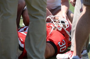
When an athlete is suspected of having a concussion, they should be removed immediately from competition. Symptoms should be monitored and the athlete should not be returned to competition until they are evaluated by a qualified medical professional. This evaluation should occur as soon as possible. The athlete should be monitored closely for several hours after a concussion. It is important to stress that both physical and mental rest speed the recovery of concussions. It is okay for the athlete to sleep and should avoid over stimulation such as video games or loud crowded activities. Athletes may need to stay out of school or have modified class schedules.
Ask your health care provider for more specific recommendations. Returning the athlete to play starts when the athlete is symptom free. It will take 3-7 days for full return to sports (depending on the sport) with an athlete gradually increasing their activity level every 24 hrs. Returning to class can occur over the same timeframe and athletes should be monitored as well for any increase or recurrence of symptoms. Activity can surface underlying concussion symptoms and athletes should be instructed to notify their coach, trainer or physician if they redevelop any symptoms during the recovery period. This process allows faster and safer return to sporting activity. Computerized neuropsychological testing is sometimes used to help monitor an athlete's progress but is never used on its own to determine a diagnosis or an athlete's readiness to return to play. There are many common misconceptions about concussive injuries.
The following are several myths about concussion:
Every athlete who sustains a hard hit must have a concussion. Although our knowledge about the forces involved in concussion is improving we still have not found a level of force that definitely causes a concussion. At times high forces do not cause an injury and relatively lower ones may. This means that we should not overact to every head impact but also need to listen to athletes who complain of concussive like symptoms after any head contact. Because there is no known force level for concussion in-helmet devices that are marketed to consumers as "concussion alarms," they are not recommended as they will likely lead to both over and under diagnosis of concussive injuries.
Better helmets and mouth guards will prevent concussions. Unfortunately there is no good scientific evidence that helmets of any type (hard shells, soft-padded or head bands) or mouth guards can prevent or reduce the risk of concussions. Hard helmets can reduce the risk of more serious head injuries (bleeding, skull fractures etc.) and should be worn in high risk sports. Mouth guards can prevent dental injuries and should be worn for sports with a high risk of these injuries. Helmet-add ons additionally are not effective in concussion prevention and using these will generally void any warranties associated with the helmet. Risk reduction may be possible in some settings with rule changes (e.g. no hitting from behind in hockey) and behavior changes (e.g. tackling technique in football).
Once you have a concussion you will always be more susceptible to having another one. While there appears to be an increased risk of recurrence in the first few weeks after a concussive injury it is unclear what factors may influence the risk of another injury in the future. Despite being a commonly held belief there is no evidence to suggest that athletes develop a decreasing force threshold after each injury. A few small studies have found the opposite....
Read more here

Friday, August 22, 2014

Researchers make functional tissue like a brain

Researchers have made a functional brain-like tissue that could have huge implications for studying neural diseases.

Researchers who created functional 3-D brain-like tissue say it could help scientists find new treatments for brain injuries and diseases and improve knowledge about normal brain function.
The tissue, which can be kept alive in the laboratory for more than two months, is structurally similar to tissue in a rat's brain. It's also functionally like brain tissue.
In early experiments with the tissue, researchers used it to study chemical and electrical changes that occur immediately after brain injury and the changes that occur in response to a drug.
The tissue was developed at Tuft University's Tissue Engineering Resource Center, which is funded by the U.S. National Institute of Biomedical Imaging and Bioengineering (NIBIB). The research is described in an article published online Aug. 11 in the Proceedings of the National Academy of Sciences.
"This work is an exceptional feat," Rosemarie Hunziker, program director of Tissue Engineering at NIBIB, said in an agency news release. "It combines a deep understand of brain physiology with a large and growing suite of bioengineering tools to create an environment that is both necessary and sufficient to mimic brain function."
This tissue offers advantages over using live animals to study brain injury, according to project leader David Kaplan, a professor of engineering at Tufts and director of the Tissue Engineering Resource Center.
In live animals, researchers can't start assessing the effects of a brain injury immediately after it occurs. That's because the animal's brain has to be dissected and prepared for experiments.
With the new 3-D brain-like tissue, "you can essentially track the tissue response to traumatic brain injury in real time," Kaplan said. "Most importantly, you can also start to track repair and what happens over longer periods of time."
The longevity of the tissue also makes it valuable for studying brain diseases and disorders.
"The fact that we can maintain this tissue for months in the lab means we can start to look at neurological diseases in ways that you can't otherwise because you need long timeframes to study some of the key brain diseases," Kaplan said.
He and his colleagues are now trying to find ways to make the tissue model even more brain-like.
Read more here

Sunday, August 17, 2014

Behavior-focused therapies and autism

This review discusses the benefits of behavior-focused therapies for children with autism.

Vanderbilt researchers this week reported updated findings regarding the benefits of behavior-focused therapies for children with autism spectrum disorder (ASD).
The review, conducted by the Agency for Healthcare Research and Quality (AHRQ)-funded Vanderbilt Evidence-based Practice Center (EPC), updates a prior systematic review of interventions for children (up to age 12) with a focus on recent studies of behavioral interventions.
Study authors said the quality of research studies has improved dramatically since AHRQ's 2011 review of studies on ASD, when authors reported that there were significant gaps in research available to document the benefits of treatments.
The updated review examined published evidence regarding the effectiveness of early intensive behavioral and developmental interventions -- interventions with a primarily behavioral approach based on applied behavioral analysis (ABA) principles and a comprehensive focus targeting multiple areas of functioning.
"We are finding more solid evidence, based on higher quality studies, that these early intensive behavioral interventions can be effective for young children on the autism spectrum, especially related to their cognitive and language skills," said lead author Amy Weitlauf, Ph.D., assistant professor of Pediatrics and a Vanderbilt Kennedy Center investigator. "But the individual response to these treatments often varies from child to child."
"We are also finding evidence that some of these targeted interventions, especially related to cognitive treatments for anxiety disorders, are also very effective for many, many children. Again, responses vary substantially and there are some children for whom these treatments have not yet been studied. So there is lots of promising evidence that these interventions are helpful, but we definitely need more research on which kids the treatments are more helpful for over time," Weitlauf said.
In previous decades ASD was often thought to be almost untreatable, said senior author Zachary Warren, Ph.D., director of the Vanderbilt Kennedy Center's Treatment and Research Institute for Autism Spectrum Disorders (TRIAD).
But researchers, clinicians and parents have increasingly documented impressive improvements in early cognitive, language and educational outcomes for some children with ASD receiving intervention.
Research results were not as strong in terms of improving broad developmental outcomes of children with ASD when interventions focused on training parents to use behaviorally based approaches with their children, but the interventions did show positive effects on parenting behaviors, on interactions between parents and children and on communications skills for some children.
"Given the potential for interventions to powerfully improve children's quality of life, in combination with the significant costs and resources often associated with treatment, it is not surprising that many groups -- parents, providers, policymakers, insurance providers -- are searching for an enhanced understanding of which interventions work the best for children with ASD," said Warren, associate professor of Pediatrics, Psychiatry and Special Education.
Read more here

Saturday, April 05, 2014

One stop shop for a scholarly review of state-of-the-art research on autism in children

This article reviews the recent research that has been done on autism in 6 main areas.

Recent years have seen exciting progress in key areas of research on autism spectrum disorders (ASD): from possible genetic causes, to effective treatments for common symptoms and clinical problems, to promoting success for young people with ASD entering college. Updates on these and other advances in ASD research are presented in the March special issue of Harvard Review of Psychiatry.
"Autism is one of the most challenging disorders to treat and the public health concerns associated with the disorder are numerous due to its burden on the individual, on the family and on society," write guest editors Drs Jean A. Frazier of University of Massachusetts Medical School and Christopher J. McDougle of Harvard Medical School. The special issue provides a timely update on research into the causes, important clinical issues, and evidence-based treatments for ASD.
Updates on ASD Research in Six Key Areas
Leading experts provide state-of-the-art reviews on six topics: the genetics of ASD; the use of psychoactive drugs; symptoms of special clinical problems, including obesity, gastrointestinal problems, and sensory issues; and transitioning to college.
Genetics. Over the last few years, there have been "unprecedented advances" in understanding the genetic causes of ASD. Hundreds of genes conferring varying degrees of ASD risk have been identified to date. Many of these genes also appear to be risk factors for related neurodevelopmental disorders and psychiatric problems. While many unanswered questions remain, it may soon be possible to make specific genetic diagnoses in children with ASD.
Psychoactive medications. Despite limited evidence, psychotropic drugs are widely used to manage behavior problems and mental health disorders in children with ASD. For medication classes -- including antidepressants and stimulants -- effectiveness may differ for youth versus adults with ASD. New treatments affecting specific neurotransmitters and the hormone oxytocin are under development, and may help in targeting the "core symptoms" of ASD.
Obesity. Obesity is a common problem with a major impact on the health of children with ASD. Some ASD-related genes may also promote obesity; the same is true for antipsychotic drugs used to help manage behavior problems. Other contributing factors may include sleep disorders and barriers to getting enough exercise. Childhood obesity and related health issues may be a "significant threat" to the health and quality of life of children with ASD.
Gastrointestinal issues. Children with ASD also have high rates of gastrointestinal symptoms and disorders. Some genes linked to ASD may also play a role in gastrointestinal disturbances, with a possible link to immune system dysfunction. There's also emerging evidence of a potential "gut-brain" connection, with gastrointestinal dysfunction contributing to the development or severity of ASD symptoms.
Sensory symptoms. Children with ASD have various abnormalities of sensory function, including both over- and under-responsivity as well as "sensory seeking behavior." Although the neurobiology of these sensory symptoms remains unclear, some researchers suggest they are related to known abnormalities of brain structure and function. Recent studies show that sensory symptoms are related to other ASD-related symptoms and behaviors; more research is needed to demonstrate the effectiveness of "sensory integration therapy" and other occupational therapy approaches.
Preparing for college. The final article highlights the need for new approaches to meeting the needs of "high-functioning" ASD patients entering college. Students "on the spectrum" transitioning to college are at risk of both academic and social problems, and may benefit from accommodation and supports. Based on a growing body of research, a set of recommendations for developing more effective transition plans for children with ASD are proposed.
Along with the editors of Harvard Review of Psychiatry, Drs Frazier and McDougal hope their special issue will provide a useful update for clinicians caring for the growing numbers of individuals and families living with ASD. They conclude, "Clearly, more research is needed on every level for the field to help support and treat individuals on the spectrum so that they can optimize their developmental trajectory and as adults become integral members of our work force."
The journal can be found online at:http://journals.lww.com/hrpjournal/Pages/default.aspx

Monday, December 30, 2013

How Botox can help with Cluster Headaches

This article discusses research that is currently being done to turn Botox into a treatment for cluster headaches.

Botox is best known as a facial wrinkle remover, but if all goes well with the research, someday soon it may be a treatment option for cluster headache. At least that is the hope of a team of scientists at the Norwegian University of Science and Technology (NTNU).
Why Botox?
Botox (onabotulinumtoxinA) is a neurotoxin and a drug that uses a potent poison called botulinum toxin, which is derived from the bacterium Clostridium botulinum. When it is injected into facial muscles, it paralyzes them temporarily and causes wrinkles to disappear for several months.
Similarly, injections of the toxin are used to block nerve signals that cause muscles contractions in cerebral palsy and bladder spasms, to block postsurgical pain and foot pain, and to temporarily relax eye muscles in people with strabismus (misaligned eyes). Botox also is sometimes used to treat migraine.
Cluster headache and Botox
Cluster headache pain is more severe than migraine pain and can drive patients to perform desperate acts, even suicide, to avoid it. Therefore finding effective treatments for this devastating condition, which affects an estimated 53 people per 100,000 per year, is essential.
That’s why Erling Tronvik, NTNU senior consultant and researcher, along with two colleagues, are about to undertake a study of the impact of Botox on cluster headache sufferers. This team has devised a treatment device that will allow them to shoot Botox through a hole in the nasal wall into a nerve bundle located behind the sinuses.
Clinicians will use magnetic resonance imaging (MRI) scans to accurately identify the location of the nerve bundle in each patient before treatment is initiated. According to Tronvik, this unique approach should (in theory) reduce or eliminate the flow of signals in this area for three to eight months, after which time patients would need to get another treatment.
“We designed the equipment ourselves, and Botox has never been used for this anywhere else,” noted Tronvik. Soon, 10 patients will enter the first pilot study of this treatment method.
If the results of the pilot study are positive, the team plans to enroll 30 to 40 patients who suffer with cluster headache and about 80 migraineurs as well.
Are there any side effects? Tronvik explained that use of MRI is a highly accurate way to locate the exact spot to inject the Botox. However, he also noted that if the toxin were to slightly miss the mark, patients could experience a weakened ability to chew or temporary double vision.
In the meantime, there’s some good news regarding chronic migraine. Botox has been shown to be helpful in relieving chronic migraine, as seen in the results of the PREEMPT (Research Evaluating Migraine Prophylaxis Therapy) clinical program.
In that study, nearly 70 percent of patients treated with Botox experienced at least a 50 percent reduction in the frequency of headache days. Other research has shown Botox to be effective as preventive treatment for chronic migraine and to provide a reduction in severity and intensity of pain as well as the number of days with disability.
If you suffer with cluster headache, a new effective treatment can’t come soon enough. Hopefully Botox or another option in the pipeline will prove beneficial in the near future.
Study References
Alvaro-Gonzalez LC et al. Botulinum toxin A in chronic refractory migraine: premarketing experience. Revista de Neurologia 2012 Oct 1; 55(7): 385-91
Aurora SK et al. OnabotulinumtoxinA for chronic migraine: efficacy, safety, and tolerability in patients who received all five treatment cycles in the PREEMPT clinical program. Acta Neurologica Scandinavica 2014 Jan; 129(1): 61-70
Read more here

NIH and NFL will research long-term effects of concussions

This article outlines research that will be done on the long-term effects and consequences of concussions. This research will be done as a partnership between the National Institutes of Health (NIH) and the National Football League (NFL).

The U.S. National Institutes of Health is teaming up with the National Football League on research into the long-term effects of repeated head injuries and improving concussion diagnosis.
The projects will be supported largely through a $30 million donation made last year to the Foundation for the National Institutes of Health by the NFL, which is wrestling with the issue of concussions and their impact on current and former players.
There's growing concern about the potential long-term effects of repeated concussions, particularly among those most at risk, including football players and other athletes and members of the military.
Current tests can't reliably diagnosis concussion. And there's no way to predict which patients will recover quickly, suffer long-term symptoms or develop a progressive brain disease called chronic traumatic encephalopathy (CTE), according to an NIH press statement released Monday.
"We need to be able to predict which patterns of injury are rapidly reversible and which are not. This program will help researchers get closer to answering some of the important questions about concussion for our youth who play sports and their parents," Story Landis, director of the National Institute of Neurological Disorders and Stroke (NINDS), said in the news release.
Two of the projects will receive $6 million each and will focus on determining the extent of long-term changes that occur in the brain years after a head injury or after numerous concussions. They will involve researchers from NINDS, the National Institute of Child Health and Human Development and academic medical centers.
One of the projects will attempt to define a clear set of criteria for various stages of CTE. It will also seek to distinguish it from Alzheimer's disease, amyotrophic lateral sclerosis (Lou Gehrig's disease) and other degenerative brain diseases that as of now can only be determined in brain tissue samples collected after death. The objective is to find medical signs of CTE that might eventually be used to diagnose the illness in living people.
The other project will seek to identify the long-term effects of mild, moderate and severe traumatic brain injury (TBI) and compare them with features of CTE. The goal is to identify signs that could be used to diagnose brain degeneration linked to traumatic brain injury in patients.
While the two projects focus on different aspects of traumatic brain injury, "their combined results promise to answer critical questions about the chronic effects of single versus repetitive injuries on the brain, how repetitive TBI (traumatic brain injury) might lead to CTE, how commonly these changes occur in an adult population, and how CTE relates to neurodegenerative disorders like Alzheimer's disease," Landis said.
Six other pilot projects will receive a total of just over $2 million and last up to two years. They will concentrate on improving the diagnosis of concussions and identifying potential medical signs that can be used to assess a patient's recovery. If the early results are promising, these projects may form the basis of more extensive research, the news release said.

Tuesday, April 16, 2013

Study: Long-term effects of concussions tested by university student

University students in Georgia are leading an interesting study on the long-term effects of concussions.


From up in the stands on a Friday night, high school football spectators hear it — the grunt of an athlete, the whistle of a referee, the crack of a helmet against another helmet.
Down on the field, the player feels it. Every time he gets tackled to the ground, the player knocks his head against the inside of his helmet. Sometimes he’s fine, but, in less fortunate occasions, he sustains a concussion.
Doug Terry, a third-year Ph.D. student in clinical psychology, and others in the University of Georgia psychology department are studying the effects of those concussions in an experiment he said they hope to finish by the time August rolls around.
“This is a global health problem,” the Long Island, N.Y., native said. “The most common way to get a concussion is a sports-related injury. I’ve worked at Children’s Healthcare of Atlanta and now concussions are becoming an increasingly big concern. There’s almost panic in parents who have children — and children themselves, these days — related to if you get a concussion, what that’s going to mean later in life.”
Their research is based on a study released last fall by the National Institute for Occupational Safety and Health, which found that NFL players are at a higher risk of dying from complications with neurodegenerative diseases than the average population.
Concussions, Terry said, are a probable link to deaths caused by neurodegenerative diseases such as Alzheimer’s and amyotrophic lateral sclerosis.
“What we’re seeing in the NFL players is that they might have gotten a concussion and they recovered,” he said. “But then, 40 years later, they’re getting Alzheimer’s at higher rates or they have memory issues more so than people without concussions.”
So, in conjunction with Stephen Miller, a professor in the department of psychology and the director of the Bio-Imaging Research Center, Terry decided to test the NIOSH conditions in a way that would make the results less exclusive.
“How many concussions is too many?” Terry said. “That’s why I’m looking at a population that typically has fewer concussions than NFL players.”
He revamped his initial experiment so that there was a greater time lapse between sustaining a concussion and the after effects of the injury. This time, he wanted to test men in the age range of 40 to 65 who had, while playing football in high school (and not after), received a concussion.
The important part of the injury history, Terry said, is that the concussion had to be a result of playing football. Anyone who might have obtained a head injury in a car accident or a fall would not fit into the experiment because of the amount of force put on the brain.
“Picture yourself on a football field, getting tackled by somebody and hitting the ground and getting a concussion that way, versus going 50 miles an hour in a car and hitting a wall or another car,” he said. “In one, you’re getting whiplash. You’re going forward and then you’re going back in a car. That type of injury is different than if you’re getting tackled and your head just hits the floor and stops.”
The experiment
Last year, Terry tested the effects of concussions on UGA rugby players. The results were not surprising, he said.
“The guys were still in college, so they had had concussions and recovered from those concussions for at least six months,” he said. “We scanned them and did all of these different memory tasks. In that study, the young rugby players didn’t have any issues related to the concussions they had sustained.”
Miller, Terry’s mentor, said there is a possibility concussions at a young age can cause some cognitive changes that aren’t detected because of the injured person’s “top health.”
“If subtle damage or change occurred, over time it may have either an effect that can no longer be overcome by youth and high health, or an effect that worsens over time, to the point that 20 or more years later it begins to show up,” Miller said. “Of course, a third possibility is that having a series of concussions when you are young has no effect on you when in your 50s and 60s.”
To test those conclusions, Terry and company have combined memory tests and functional magnetic resonance imaging scans. The tasks include tests of working memory, which Terry said was the ability to concentrate on and do several things at once.
“When someone does a working memory task, their brain will use more resources,” he said. “After you get a concussion, your brain uses more oxygen, more neurons are firing and more glucose is used up by the brain. Your brain has to work harder to do the same thing. There’s evidence of that in functional MRI directly after a concussion.”
Patrick Curry, a second-year athletic training master’s student from Durham, Conn., is a UGA athletic trainer who has seen all the concussions sustained by University athletes in the last two years. He introduced Terry to a system, the “NeuroCom Balance Manager,” that can test the effects of concussions.
The benefits of using that machine in addition to the psychological tests Terry runs, Curry said, is that it has more capability than a general assessment.
“It’s an objective measure of balance that uses two force plates to measure your center pressure,” Curry said. “It tests all your systems of balance. It can see if balance is impaired many years after a concussion occurs.”
What Terry said he hopes to find in the experiment is the kind of effect, if any, a concussion has on an individual years after the injury.
Curry said the experiment’s results could have a major effect on the way teams handle concussions.
“The biggest thing we’re looking at right now and the biggest concern in sports medicine is the long term effects of concussions,” he said. “This experiment could be a great way to look at it. It could change how we manage concussions acutely. It could change our return to play criteria after someone sustains a concussion.”
Read more here

Friday, April 05, 2013

Genetic discovery for common form of epilepsy

Research from Australia has found a gene indicating one of the most common forms of epilepsy. This could have major implications for future treatment options.


An Australian-led international research team has discovered a gene associated with the most common form of epilepsy, a discovery that will help with diagnosis and new treatments.

Through genetic counselling, it will help people plan a family, lead researcher Professor Ingrid Scheffer of the University of Melbourne said.

Two per cent of people have epilepsy and most do not know the cause of their condition. The research will help some of those with the most common form, focal epilepsy, discover the underlying cause.

Professor Scheffer said a gene test would help in cases where everything else in the brain looked normal. ''It will give you a cause. That has important implications in terms of genetic counseling and managing the risk to your own offspring.''

She said a small proportion of people with the gene also had psychiatric or autism-spectrum disorders.

''Therefore genetic counselling is even more important. Knowing the gene means people can go forward and get pregnant and have proper medical assistance to ensure their baby does not have the disorder.''

Professor Scheffer said 90 families took part in the study, which was conducted in partnership with Associate Professor Leanne Dibbens of the University of South Australia.

Scientists in Europe and Canada also worked on the research, which is published in the journal Nature Genetics.

Read more here

Saturday, March 30, 2013

Zebrafish research promises new epilepsy treatment

Research on zebrafish shows compounds that help suppress seizures showing promise for future epilepsy treatment.


The prospect of developing new treatments for epilepsy sufferers has been given a boost by a pioneering discovery at a leading international centre of research into human disease in the University of Sheffield.
Researchers at the University’s Medical Research Council Centre for Developmental and Biomedical Genetics (CDBG), in the Department of Biomedical Science, screened a collection of 2,000 biologically active compounds to identify molecules that suppressed epileptic seizures in two day old epileptic zebrafish.
Within this collection, 46 compounds – including some which are used to treat infectious, psychiatric and inflammatory disorders – were found to exhibit anticonvulsant activity and could represent starting points for the development of new drugs for treating epilepsy.
Approximately one out of every 140 people in the UK has epilepsy – more than 400,000 people – of which about 30 per cent do not respond favourably to the available anti-epileptic drugs.
Consequently, many patients live with the disruptive and often devastating effects of untreatable seizures in their daily lives, whilst other patients who receive medication for their seizures experience side-effects that can result from taking some of these drugs.
The University of Sheffield team’s innovative approach to identifying small molecules with potential as anti-epileptic therapies offers new prospects of reducing the burden of suffering from this devastating illness.
Dr Vincent Cunliffe of the University of Sheffield’s Department of Biomedical Science, who led the project, said: “We took advantage of a unique set of features of the zebrafish to look for new anticonvulsant agents within a library of many different types of compounds with a wide range of known biological activities.
“We found that a small number of them had previously-unknown anti-convulsant effects. Some of the identified compounds already have a variety of different medical uses in treating conditions such as fungal infections, as well as psychiatric and inflammatory disorders.”
The research, published in the journal Disease Models & Mechanisms, suggests that some of these existing drugs could be re-purposed for treatment of epilepsy.
Nerve cells communicate with one another by passing electrical impulses along their lengths, leading to the release of a variety of chemical signals known as neurotransmitters at nerve endings, which may then stimulate or inhibit neighboring cells.
Epileptic seizures occur as a result of imbalances in the types of neurotransmitters produced within the brain, causing the simultaneous activation of abnormally large numbers of nerve cells, some of which may then stimulate body muscles to contract vigorously, resulting in convulsions.
Observing these processes at the level of individual nerve cells and molecules is especially difficult because the brains of mammals such as humans and mice, are so large, complex and relatively inaccessible.
However, the three milimetre-long, microscopic zebrafish larva develops rapidly, independently of its parents, and it is structurally simple, transparent and accessible, which allows the behaviours of nerve cells within the brain to be easily viewed in a remarkable level of detail. To study the effects of drugs on the zebrafish brain, they are simply diluted into the water in which the zebrafish develop, which then allows them to be readily absorbed by the body.
Dr Cunliffe added: “The zebrafish is proving to be a remarkably powerful in vivo system for gene function analysis and drug discovery. Over the last ten years our zebrafish research has helped us to understand how the nervous system is built and how faults in this construction process may cause neurological and psychiatric diseases.
“Three years ago we began to explore the usefulness of the zebrafish for drug discovery and we have been surprised by the success we have had in a relatively short period of time.”
More traditional approaches to identifying and developing new pharmaceuticals are slower and more costly, so adopting the zebrafish – a small tropical fish of the minnow family – for this type of research, could help to shorten the timescales and reduce the overall costs of drug development.
Read more here


New technologies utilized for epilepsy treatment

This article claims that developing new technologies is the best way to treat epilepsy. It also goes over a few of the technologies currently utilized for epilepsy treatment.

Speaking in the lead up to Purple Day for Epilepsy Awareness (Tuesday 26 March), geneticist Professor Jozef Gecz says advances in DNA sequencing have been a huge leap forward in understanding epilepsy. 

This, combined with the use of stem cells in laboratory research, will lead to further advances in epilepsy treatment, he says. 

However, he cautions that the same technology has also helped to reveal that epilepsy is a far more complex condition than previously thought. 

"Scientists used to believe that epilepsy was just one condition, possibly with one main cause. But now we know it is a very complex series of neurological disorders – it is many epilepsies, instead of just one epilepsy, with multiple causes and various symptoms," says Professor Gecz, from the University of Adelaide's School of Paediatrics and Reproductive Health. 

Epilepsy is common, with up to 3% of the Australian population experiencing epilepsy at some stage in their lives. Genetic and environmental factors, and trauma, can all play a role in the development of epilepsy. Most (but not all) forms cause sufferers to experience seizures, which vary in severity. 

Research in Adelaide has played an important role in the understanding of epilepsy in recent years. 

"It's really thanks to the pioneering work of Dr John Mulley (Women's and Children's Hospital and University of Adelaide), who discovered the first gene for idiopathic epilepsy almost 20 years ago. Since then, almost 40 idiopathic epilepsy genes have been discovered, many of them by researchers here in Adelaide," Professor Gecz says. 

"There are more than 300 genes known today in which DNA mutations can give rise to some form of epilepsy, in addition to other problems like intellectual disability, autism or psychiatric problems. 

"Thanks to genetic sequencing technology, in most cases we are now able to solve the mystery about what kind of epilepsy a patient has, and we can do this very quickly, very accurately, and cost effectively. 

"Molecular diagnosis is making a huge impact on treatment – it's really taken off in the last few years, and it has the potential to be even more effectively used in the future. Clinicians can now be guided by genetic information when considering treatment of patients with specific epilepsies." 

Professor Gecz and colleagues are currently involved in a major national study of epilepsy, with his lab focusing on the "genetic architecture" of the condition.

Read more here

Sunday, March 17, 2013

Canadian Perspectives - Current Epilepsy Research & Facts

This article reviews a Canadian perspective on some advances in epilepsy research and lists some interesting epilepsy facts.

Many facts are relevant here in the US too. Many people do not repsond to current medications.

 JR


A biomarker for epilepsy
The use of electroencephalography (EEG) for the study and treatment of epilepsy was pioneered at The Neuro. New research demonstrates the value of using EEG to monitor high-frequency oscillations (HFOs) in a patient’s brain. The research shows that information from HFOs can be used as a valuable biomarker, which is an indicator of a diseased state and aids epilepsy diagnosis.  HFOs also appear to provide a better indication of the specific brain tissue that surgeons must remove to prevent a patient’s seizures.  Furthermore, the link between HFOs and the source of epileptic seizures strongly suggests that further study of HFOs could reveal information about the genesis of epilepsy, especially in children.  This research was led by The Neuro’s Jean Gotman, in collaboration with colleagues, Francois Dubeau and Rina Zelmann.

Discovering epileptic networks in the brain
Recent research at The Neuro suggests that different epileptic syndromes can lead to the formation of unique, widespread neural networks.  These networks are related to discharges in the periods between a person’s epileptic seizures (interictal epilepsy discharge, or IED).  Researchers simultaneously used EEG and functional magnetic resonance imaging (fMRI) to examine patients with different syndromes---frontal lobe epilepsy, posterior quadrant epilepsy and temporal lobe epilepsy.   The conclusion was that changes in the brain’s metabolism during IEDs might cause more widespread brain dysfunction than would be indicated by EEG results of discharges monitored only at the level of the scalp. The research was published by lead author Jean Gotman and his colleagues at The Neuro, Firas Fahoum, Renaud Lopes, Francesca Pittau, and Francois Dubeau.

EPILEPSY FACTS:
  • Epilepsy is a neurological disorder characterized by a sudden, brief change in the brain, expressed as a seizure.
  • A seizure can appear as a convulsion lasting a few seconds or a few minutes, but a seizure can also be not more than a brief stare, an unusual movement of the body or a change in awareness.
  • Epilepsy affects many Canadians.  According to Epilepsy Canada, approximately 0.6% of Canadians have epilepsy. This includes people who take anticonvulsant drugs or who had a seizure within the past five years.
  • 30% of new Canadian cases each year are among children.  In about half of child cases,seizures eventually disappear.
  • Epilepsy is a result of different causes: malformations during brain development, a head injury that causes scarring to the brain tissue, high fever and prolonged convulsions during early childhood, trauma at birth, a stroke or tumour.
  • 1 out of 3 patients cannot control seizures solely by using available medications. For these patients, surgical removal of the brain tissue causing seizures is the only known effective treatment for controlling seizures and improving quality of life.
  • Seizures can be triggered by outside events such as strobe lights, or by a person’s state of health---fatigue, illness, allergies, hunger, emotional stress.

Friday, January 11, 2013

Genetic Link Fount Between Migraine and Epilepsy

Research has found medical evidence of a genetic link between migraine headaches and epilepsy. The research indicates that a strong family history of epilepsy increases the likelihood that an individual will have migraines.

New research reveals a shared genetic susceptibility to epilepsy and migraine. Findings published in Epilepsia, a journal of the International League Against Epilepsy (ILAE), indicate that having a strong family history of seizure disorders increases the chance of having migraine with aura (MA).

Medical evidence has established that migraine and epilepsy often co-occur in patients; this co-occurrence is called "comorbidity." Previous studies have found that people with epilepsy are substantially more likely than the general population to have migraine headache. However, it is not clear whether that comorbidity results from a shared genetic cause.
"Epilepsy and migraine are each individually influenced by genetic factors," explains lead author Dr. Melodie Winawer from Columbia University Medical Center in New York. "Our study is the first to confirm a shared genetic susceptibility to epilepsy and migraine in a large population of patients with common forms of epilepsy."
For the present study, Dr. Winawer and colleagues analyzed data collected from participants in the Epilepsy Phenome/Genome Project (EPGP) -- a genetic study of epilepsy patients and families from 27 clinical centers in the U.S., Canada, Argentina, Australia, and New Zealand. The study examined one aspect of EPGP: sibling and parent-child pairs with focal epilepsy or generalized epilepsy of unknown cause. Most people with epilepsy have no family members affected with epilepsy. EPGP was designed to look at those rare families with more than one individual with epilepsy, in order to increase the chance of finding genetic causes of epilepsy.
Analysis of 730 participants with epilepsy from 501 families demonstrated that the prevalence of MA -- when additional symptoms, such as blind spots or flashing lights, occur prior to the headache pain -- was substantially increased when there were several individuals in the family with seizure disorders. EPGP study participants with epilepsy who had three or more additional close relatives with a seizure disorder were more than twice as likely to experience MA than patients from families with fewer individuals with seizures. In other words, the stronger the genetic effect on epilepsy in the family, the higher the rates of MA. This result provides evidence that a gene or genes exist that cause both epilepsy and migraine.
Identification of genetic contributions to the comorbidity of epilepsy with other disorders, like migraine, has implications for epilepsy patients. Prior research has shown that coexisting conditions impact the quality of life, treatment success, and mortality of epilepsy patients, with some experts suggesting that these comorbidities may have a greater impact on patients than the seizures themselves. In fact, comorbid conditions are emphasized in the National Institutes of Health Epilepsy Research Benchmarks and in a recent report on epilepsy from the Institute of Medicine.
"Our study demonstrates a strong genetic basis for migraine and epilepsy, because the rate of migraine is increased only in people who have close (rather than distant) relatives with epilepsy and only when three or more family members are affected," concludes Dr. Winawer. "Further investigation of the genetics of groups of comorbid disorders and epilepsy will help to improve the diagnosis and treatment of these comorbidities, and enhance the quality of life for those with epilepsy."
Read more here

Saturday, December 15, 2012

Severity of Autism and Symptoms May Be Due to Fear

Research on autism shows that the severity of autism diagnoses may be due to anxiety. This could also explain some of the other symptoms and severity of autism's disability.  For many children with autism "My way IS the highway." 

Anxiety is disabling for families and impedes developmental progress.

JR

Most people know when to be afraid and when it's okay to calm down.

But new research on autism shows that children with the diagnosis struggle to let go of old, outdated fears. Even more significantly, the Brigham Young University study found that this rigid fearfulness is linked to the severity of classic symptoms of autism, such as repeated movements and resistance to change.
For parents and others who work with children diagnosed with autism, the new research highlights the need to help children make emotional transitions -- particularly when dealing with their fears.
"People with autism likely don't experience or understand their world in the same way we do," said Mikle South, a psychology professor at BYU and lead author of the study. "Since they can't change the rules in their brain, and often don't know what to expect from their environment, we need to help them plan ahead for what to expect."
In their study, South and two of his undergraduate neuroscience students -- Tiffani Newton and Paul Chamberlain -- recruited 30 children diagnosed with autism and 29 without to participate in an experiment. After seeing a visual cue like a yellow card, the participants would feel a harmless but surprising puff of air under their chins.
Part-way through the experiment, the conditions changed so that a different color preceded the puff of air. The researchers measured participants' skin response to see if their nervous system noticed the switch and knew what was coming.
"Typical kids learn quickly to anticipate based on the new color instead of the old one," South said. "It takes a lot longer for children with autism to learn to make the change."
The amount of time it took to extinguish the original fear correlated with the severity of hallmark symptoms of autism.
"We see a strong connection between anxiety and the repetitive behaviors," South said. "We're linking symptoms used to diagnose autism with emotion difficulties not usually considered as a classic symptom of autism."
The persistence of needless fears is detrimental to physical health. The elevated hormone levels that aid us in an actual fight or flight scenario will cause damage to the brain and the body if sustained over time.
And the families who participate in social skills groups organized by South and his students can relate to the new findings.
"In talking to parents, we hear that living with classic symptoms of autism is one thing, but dealing with their children's worries all the time is the greater challenge," South said. "It may not be an entirely separate direction to study their anxiety because it now appears to be related."
The complete study appears in the journal Autism Research.The project began when Newton received a university grant to conduct research with a faculty mentor. After graduating with a degree in neuroscience, she began working at a clinic in Michigan. Chamberlain is finishing his senior year at BYU and is currently interviewing with medical schools.
Read more here

Tuesday, September 18, 2012

Researchers show scientific proof of deficiencies in football helmets

Researchers have produced biomechanical tests to show how current football helmets do not protect football helmets from concussions.

Researchers at BRAINS, Inc. have conducted biomechanical tests revealing the deficiency of current football helmet designs in protecting players from brain injury, particularly concussion.

Historically, helmet effectiveness has been measured through drop-tests, using a device approved by the National Operating Committee on Standards for Athletic Equipment (NOCSAE). The result is helmets that are optimized against skull fractures, bruising, and other focal effects. 

“We modified the standard test device to consider rotationalacceleration in addition to conventional linear impact measures” explains John Lloyd, PhD.

Biomechanical researchers have long understood that angular forces can cause serious brain damage including concussion, axonal injury, and hemorrhages.

Using proprietary miniature sensors to measure concussion risk at the center of the brain, BRAINS researchers completed more than 330 tests across ten popular helmet brands. The team concluded that while these helmets provide excellent protection from linear impacts – those leading to bruising and skull fracture – they offer little or no protection against angular acceleration, a dangerous source of brain injury and encephalopathy.

The graph in the slideshow above shows percent reduction in linear impact acceleration, Head Injury Criterion (HIC), and angular acceleration provided by the different football helmets, compared to the same impact with no helmet. Note that all helmets provide considerable protection from skull fracture (blue) and focal brain impact (green), but are far less effective at reducing risk of diffuse brain injury and concussion and encephalopathy (red). In fact, some helmet designs offer no significant protection from concussion — and those that offer the least protection are among the most popular on the field.

The table, also in the slideshow above, presents a ranking of the more popular football helmets, from best to worst, based on their combined protection from skull fracture, focal brain impact and diffuse brain injury.

Protection against concussion and axonal injury is especially important for young players, including peewee, high school, and college participants, whose still-developing brains are more susceptible to the lasting effects of encephalopathy. Therefore, the need to develop headgear to protect susceptible individuals from life-changing brain damage is paramount.

Consistent with their innovative approach to meeting the challenges of brain trauma, combined with 20+ years of experience in biomechanics, and neurophysiology, BRAINS researchers have investigated several new technologies to measure and reduce the debilitating effects of concussion in football players. The team is poised to integrate their new technology into helmet design – a paradigm-shift in helmet construction – and bring to market a more comprehensive form of head gear to defend against catastrophic brain injuries while also mitigating linear forces associated with impact.

Read more here

Saturday, September 15, 2012

Researchers find more Autism Genes


Its news like this that keeps me going. Every day, I meet parents who have given up on neurology.  Even in the last 5 years, our tools for diagnosis and treatment have changed. With advances in genetics more news will come. - JR 

Researchers at UCSD found a gene that indicated autism associated with epilepsy.

A genetic cause for a rare form of epilepsy-associated autism has been identified by UC San Diego and Yale scientists.
Moreover, symptoms of the newly discovered form have been reversed in mouse models by altering diet. This gives rise to the possibility that similar treatment might help people, the researchers said. The study was published online Thursday in the journal Science
Researchers led by Gaia Novarino and Joseph G. Gleeson of UCSD studied two families, one of Egyptian descent and another of Turkish origin. They examined the genome of patients and healthy relatives for exons, gene sequences that code for proteins. The researchers found that patients shared an exon mutation on a gene called BCKDK. The mutant gene is recessive, meaning that it must be inherited from both mother and father to manifest.
Moreover, the researchers found that the mutation caused patients to produce abnormally low levels of certain types of amino acids, the building blocks of proteins. They were able to boost levels of these amino acids to normal with a nutritional supplement from a health food store. Research is now ongoing as to whether this supplementation will reduce symptoms of epilepsy and autism in these patients.
Those who might be helped are only a small fraction of people with autism, Novarino said in an Tuesday interview. Those without the metabolic defect wouldn't benefit from the supplementation.
The study illustrates how scientists have become more sophisticated in using knowledge of the human genome to crack the puzzle of previously intractable diseases. The genome is the complete set of hereditary information encoded in DNA.
Narrowing the search
The vast majority of DNA does not code for proteins, the body's workhorse molecules. This "non-coding" DNA was ignored in the new method of DNA analysis, called "whole exome" sequencing, which looks only at the exons. An advantage of whole exome sequencing is that it focuses exclusively on proteins, which are altered or missing in genetic diseases.
Whole exome sequencing can find previously undiscovered genetic diseases, according to another study performed by some of the same UCSD researchers. They examined 118 patients diagnosed with neurological disorders who had no known genetic disease causes. In addition to the newly discovered genetic causes, in about 10 percent of cases the researchers even found a known disease-causing gene that had previously escaped detection.
That study was published in June in Science Translational Medicine, a journal devoted to getting research discoveries into the hands of doctors more quickly.
The new study is part of the same project of applying exome research to diseases, Novarino said.
Genetic knockout
In the study, the researchers produced genetically engineered "knockout mice" in which the BCKDK gene was inactivated. These mice experienced epileptic seizures, tremors, hind limb clasping and other symptoms of neurological disorders. Their brains were found to be deficient in certain chemicals called branched chain amino acids.
Seizures and hind limb clasping were "completely abolished" within a week when mice were given diets rich in these nutrients, the study said.
The researchers had previously examined neural cells of patients and unaffected family members. The neural cells were made from induced pluripotent stem cells, produced from skin cells and turned into neurons. However, the researchers couldn't find any differences in the cell cultures. That's when they turned to studying the effect of diet on whole knockout and healthy mice.
Novarino said it's too early to tell if BCAA supplementation is helping the human patients in the study.
The supplements have not caused any side effects in the patients, nor did they in the mouse, Novarino said.
The complete list of authors, including colleagues in Turkey, Egypt and Libya, can be found at the end of the press release. Funders of the study include the National Institutes of Health, the Center for Inherited Disease Research, and the Simons Foundation Research Initiative.
Read more here