Showing posts with label eyes. Show all posts

Wednesday, February 11, 2015

Eye tracking to help determine concussion severity

Using an eye-tracking method may help spot concussions and help determine their severity.
A new eye-tracking method might help determine the severity of concussions, researchers report.They said the simple approach can be used in emergency departments and, perhaps one day, on the sidelines at sporting events.
"Concussion is a condition that has been plagued by the lack of an objective diagnostic tool, which in turn has helped drive confusion and fears among those affected and their families," said lead investigator Dr. Uzma Samadani. She is an assistant professor in the departments of neurosurgery, neuroscience and physiology at NYU Langone Medical Center in New York City.
"Our new eye-tracking methodology may be the missing piece to help better diagnose concussion severity, enable testing of diagnostics and therapeutics, and help assess recovery, such as when a patient can safely return to work following a head injury," she explained in an NYU news release.
According to researchers, it's believed that up to 90 percent of patients with concussions or blast injuries have eye movement problems. But the current method of assessing eye movement is asking a patient to track a doctor's finger, Samadani said.
The new method was originally developed by Samadani and her colleagues to assess eye movement in U.S. military personnel believed to have concussion or other types of brain injuries.
The researchers compared 75 trauma injury patients and a control group of 64 healthy people. The movements of the participants' pupils were tracked while they watched a music video for a few minutes.
Thirteen trauma patients who hit their heads and had CT scans showing new brain damage, and 39 trauma patients who hit their heads and had normal CT scans, were much less able to coordinate their eye movements than trauma patients who hadn't hit their heads and those in the control group.
The more severe the concussion, the worse a patient's eye movement problems, according to the study. Results were published online Jan. 29 in the Journal of Neurotrauma.
Dr. M. Sean Grady, chair of the neurosurgery department at the University of Pennsylvania's Perelman School of Medicine in Philadelphia, said, "The importance of this study is that it establishes a reliable test and a 'biological' marker for detecting concussion." He was not involved in the study.
"Since concussion can occur without loss of consciousness, this can be particularly important in sideline evaluations in athletics or in military settings where individuals are highly motivated to return to activity and may minimize their symptoms. More work is needed to establish its sensitivity and specificity, but it is very promising," Grady said.
Read more here

Friday, August 22, 2014

ADHD diagnosis by involuntary eye movements

A study shows that ADHD can accurately be diagnosed by a person's involuntary eye movements.

Attention Deficit Hyperactivity Disorder (ADHD) is the most commonly diagnosed -- and misdiagnosed -- behavioral disorder in children in America, according to the Centers for Disease Control and Prevention. Unfortunately, there are currently no reliable physiological markers to diagnose ADHD. Doctors generally diagnose the disorder by recording a medical and social history of the patient and the family, discussing possible symptoms and observing the patient's behavior. But an incorrect evaluation can lead to overmedication with Ritalin (methylphenidate), which has parents everywhere concerned.
Now a new study from Tel Aviv University researchers may provide the objective tool medical professionals need to accurately diagnose ADHD. According to the research, published in Vision Research, involuntary eye movements accurately reflect the presence of ADHD, as well as the benefits of medical stimulants that are used to treat the disorder.
Keeping an eye on the eyes
Dr. Moshe Fried, Dr. Anna Sterkin, and Prof. Uri Polat of TAU's Sackler Faculty of Medicine, Dr. Tamara Wygnanski-Jaffe, Dr. Eteri Tsitsiashvili, Dr. Tamir Epstein of the Goldschleger Eye Research Institute at Sheba Medical Center, Tel Hashomer, and Dr. Yoram S. Bonneh of the University of Haifa used an eye-tracking system to monitor the involuntary eye movements of two groups of 22 adults taking an ADHD diagnostic computer test called the Test of Variables of Attention (TOVA). The exercise, which lasted 22 minutes, was repeated twice by each participant. The first group of participants, diagnosed with ADHD, initially took the test un-medicated and then took it again under the influence of methylphenidate. A second group, not diagnosed with ADHD, constituted the control group.
"We had two objectives going into this research," said Dr. Fried, who as an adult was himself diagnosed with ADHD. "The first was to provide a new diagnostic tool for ADHD, and the second was to test whether ADHD medication really works -- and we found that it does. There was a significant difference between the two groups, and between the two sets of tests taken by ADHD participants un-medicated and later medicated."
Foolproof, affordable, and accessible diagnosis
The researchers found a direct correlation between ADHD and the inability to suppress eye movement in the anticipation of visual stimuli. The research also reflected improved performance by participants taking methylphenidate, which normalized the suppression of involuntary eye movements to the average level of the control group.
"This test is affordable and accessible, rendering it a practical and foolproof tool for medical professionals," said Dr. Fried. "With other tests, you can slip up, make 'mistakes' -- intentionally or not. But our test cannot be fooled. Eye movements tracked in this test are involuntary, so they constitute a sound physiological marker of ADHD.
"Our study also reflected that methylphenidate does work. It is certainly not a placebo, as some have suggested."
The researchers are currently conducting more extensive trials on larger control groups to further explore applications of the test.
Read more here

Sunday, March 23, 2014

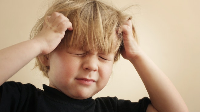
Headaches in children

This article gives a lot of information on headaches in children.

Why do children get headaches?

Headaches in children are common and usually not serious.  Causes include migraines, stress and tension headaches, trauma, sinus disease and eye problems.  Certain foods with nitrate preservatives and MSG (monosodium glutamate) can trigger headaches.  Brain tumors in children as a cause for headaches are extremely rare, and are usually associated with additional neurologic symptoms such as dizziness and lack of coordination.

How are children with headaches evaluated?

Children should have a thorough physical examination with a pediatrician.  He/she may order additional tests or refer to a neurologist.  It is helpful to keep a headache diary so that possible triggers (certain foods or activities) can be discovered.
If no obvious medical cause for the headaches is found by the pediatrician, a referral to an ophthalmologist is required to perform a complete eye exam.  The entire visual system should be examined.  This includes refractive error (the need for glasses), eye alignment, and binocularity (the ability to use both eyes together).  During this examination the pupils should be dilated.

 How do eye problems cause headaches in children?

Hyperopia (farsightedness) requires extra effort to focus clearly while reading.  This can sometimes lead to fatigue and headache.  Glasses can reduce the effort required to see clearly at near and improve the headache.
The decreased ability to pull the eyes toward each other when viewing near objects (convergence), particularly while reading, may cause headaches.  This is called convergence insufficiency, and symptoms include the doubling of images or words, blurred vision, fatigue, and headaches which worsen with prolonged reading.  Glasses are sometimes prescribed.  At home eye exercises, sometimes with the help of computer software, can help treat convergence insufficiency.  Expensive in office eye exercises are rarely indicated.
 Acute infections and inflammatory diseases of the eyes can cause headaches.  These problems are often accompanied by redness of the eye and/or eyelid and light sensitivity (photophobia).  Acute glaucoma can cause headaches, but rarely affects children.  A complete exam by an ophthalmologist can rule out these conditions.
Read more here

Tuesday, November 19, 2013

Lack of eye contact in the first month of life may indicate autism

A study claims that certain signs of autism may be seen as early as the first month of life. Specifically, lack of eye contact at that age may indicate autism in a young child.

The first signs of autism may be visible as early as the first month of a child's life, according to a study published Wednesday in the scientific journal Nature.
"These are the earliest signs of autism ever observed," says lead study author Warren Jones.
Researchers at the Marcus Autism Center in Atlanta followed 110 children from birth to age 3, at which point a diagnosis of autism was ascertained. Fifty-nine babies were considered "high risk" for developing an autism spectrum disorder (ASD) because they had siblings with autism; 51 were considered "low risk" because they did not have first, second or third-degree relatives with ASD.
Data was collected at 2, 3, 4 ,5, 6, 9, 12, 15 and 24 months of age. Each time, the children watched videos showing actresses playing the role of a caregiver. "Every baby watched the same videos, and then we could measure what was different about the responses of infants later diagnosed with autism versus infants who were typically-developing," Jones says.
Lack of eye contact is one of the red flags when it comes to autism - a group of neurodevelopmental disorders that can cause significant social, communication and behavioral challenges.
Jones, who is the director of research at Marcus Autism Center and assistant professor in the Department of Pediatrics at Emory University School of Medicine, says he really expected the children later diagnosed with autism would have diminished eye contact from birth. Instead, he and his colleagues measures how much time each baby was looking at the eyes of the caregiver in the video.
"Basically from birth, (all) babies will look more at the eye part of faces," says Jones. But at about 4 to 6 weeks, he says the attention to eyes decreases, then in typical babies picks up again at 2 months. Jones found, "in the first 6 months of life we're seeing a decline in the amount of looking at other people's eyes in children who later are diagnosed with autism."
The research suggests that a baby's initial eye contact ability may be an almost a reflex-like behavior, but then there may be a second phase of development that depends on different brain and gene systems which lead to social interaction, Jones says. That's where a typically developing child's development may differ from a child with autism.
The study authors conclude that "the observation of this decline in eye fixation - rather than outright absence - offers a promising opportunity for early intervention."
This is not something parents are going to see by just holding their baby, Jones points out.  This type of eye-tracking requires sophisticated technology that can track even the slightest movement of the eye.
"It's a very interesting study with intriguing results. " says Wendy Stone, a longtime autism researcher and director of the Research in Early Autism Detection & Intervention (READI) lab at the University of Washington. But, she adds, "many researchers in this field have not seen behaviors under 6 months to be predictive of later diagnosis."
She also cautions that babies looking at videos of their mothers are not the same as the actual stimuli created by a mom interacting with her baby. "Are these babies less interested in eyes because mouths are more interesting to look at and more attractive because there's more movements? To me that's one of the big questions," says Stone.
Dr. Max Wiznitzer, a pediatric neurologist and autism specialist at the Rainbow and Babies Children's Hospital in Cleveland, Ohio, says this new study is a continuation of previous work in babies. He says this research makes sense to him. "There's a decrease in the amount of attention to eyes as an early marker of social behavior (think of it as a primitive level of socialization)." Wiznitzer suggests the failure to establish these early social skills has ramifications later as "social behavior shifts into more sophisticated patterns."
If this research bears out, then maybe at some point a pediatric practice could track eye movements as one way to diagnose a child with autism, says Stone. "But we're really, really far away from that."
Wiznitzer says this may explain why the autism symptoms may be more apparent at 18 to 24 months, "even though 'subclinical' onset was months earlier." He also suggests these study results may offer another explanation why the measles, mumps and rubella vaccine, which isn't administered until a baby is at least 12 months old, cannot be blamed for causing autism.
Everyone agrees this research needs to be replicated in bigger studies with more children, Wiznitzer says. "The authors are correct that a replication study using a larger number (of children) is necessary. Before that time, I would not devote extensive resources towards assessing eye attention in infants or designing major intervention programs."
Jones says, "what we really want to do is create growth charts for social behavior, just like we have growth charts for charting a child's height and weight." He says these those are the kind of tools that pediatricians need and parents are looking for.
Read more here

Sunday, September 15, 2013

The best beauty tip is to get enough sleep

Sleep deprivation results in physical changes in the face to indicate tiredness. To avoid this, people need to get adequate sleep.

A small new study now confirms that your best beauty trick is to get a good night's sleep.

Findings revealed what you've probably seen in the mirror after a rough night: sleep-deprived people look more, shall we say, worn-out, with redder, more swollen eyes, darker under-eye circles, more wrinkles and droopier eyelids and mouths than their well-rested selves.

People also looked sadder when sleep-deprived than after a normal sleep, and this apparent sadness was related to looking fatigued, the researchers said.

"Since faces contain a lot of information on which humans base their interactions with each other, how fatigued a person appears may affect how others behave toward them," said Tina Sundelin, author and doctoral student in the department of psychology at Stockholm University in Stockholm, Sweden. "This is relevant not only for private social interactions, but also official ones such as with health care professionals and in public safety."

To conduct their research, Sundelin and a team from Karolinska Institute in Stockholm recruited 10 subjects, who were photographed on two separate occasions: after eight hours of normal sleep and after 31 hours of sleep deprivation. The photographs were taken in the laboratory at 2:30 pm on both occasions.

Forty participants rated the 20 facial photographs with respect to various facial cues, fatigue, and sadness.

In a separate study announced earlier this year, University Hospitals Case Medical Center in Cleveland, Ohio found that poor sleepers showed "increased signs of skin aging and slower recovery from a variety of environmental stressors," such as ultraviolet radiation, the researchers said.

If you struggle with getting quality beauty sleep, WebMD and the Mayo Clinic offer the following tips to boost your slumber:

Stick to a regular sleep schedule, even on weekends. Eat well, and avoid caffeine in the evenings or overeating before bedtime. Also try sleep accessories, such as a white noise machine or ear plugs, to block out distractions.

Exercise during the day, which can aid sleep, and try to clear your mind from too much clutter before bedtime by writing in a journal beforehand, for example.

Read more here

Sunday, September 01, 2013

Facial cues of sleep deprivation

A study shows that certain facial cues of sleep deprivation, such as distinctions with the eyes, mouth and skin, signal to others that a person is sleep deprived.

A new study finds that sleep deprivation affects facial features such as the eyes, mouth and skin, and these features function as cues of sleep loss to other people.
Results show that the faces of sleep-deprived individuals were perceived as having more hanging eyelids, redder eyes, more swollen eyes and darker circles under the eyes. Sleep deprivation also was associated with paler skin, more wrinkles or fine lines, and more droopy corners of the mouth. People also looked sadder when sleep-deprived than after normal sleep, and sadness was related to looking fatigued.
"Since faces contain a lot of information on which humans base their interactions with each other, how fatigued a person appears may affect how others behave toward them," said Tina Sundelin, MSc, lead author and doctoral student in the department of psychology at Stockholm University in Stockholm, Sweden. "This is relevant not only for private social interactions, but also official ones such as with health care professionals and in public safety."
The study, which appears in the September issue of the journal Sleep, was conducted at the Karolinska Institute in Stockholm, Sweden. Ten subjects were photographed on two separate occasions: after eight hours of normal sleep and after 31 hours of sleep deprivation. The photographs were taken in the laboratory at 2:30 p.m. on both occasions. Forty participants rated the 20 facial photographs with respect to 10 facial cues, fatigue and sadness.
According to the authors, face perception involves a specialized neuronal network and is one of the most developed visual perceptual skills in humans. Facial appearance can affect judgments of attributes such as trustworthiness, aggressiveness and competence.
Read more here

Friday, August 30, 2013

Drug uses sensors in eye to stop migraines

A new drug uses light sensors in the eye to stop from triggers for migraines.

For many migraine sufferers, bright lights are a surefire way to exacerbate their headaches. And for some night-shift workers, just a stroll through a brightly lit parking lot during the morning commute home can be enough to throw off their body's daily rhythms and make daytime sleep nearly impossible. But a new molecule that selectively blocks specialized light-sensitive receptors in the eyes could help both these groups of people, without affecting normal vision.
"It took almost ten years to find and test a molecule that fit all the properties and acted in vivo as we wanted," says senior study author Satchidananda Panda , an associate professor in Salk's Regulatory Biology Laboratory.
Scientists have known for nearly a century that humans and animals can sense light even when they can't see. Before they've opened their eyes, and even before cells that allow vision have matured, newborn mice still scurry away from bright lights, and set their sleep-wake cycles based on the patterns of light and dark throughout the day. The same is true of many blind people-though they can't see what's in front of them, their bodies still follow daily circadian rhythms, and the pupils of their eyes constrict in response to light.
More than ten years ago, Panda's lab group discovered that melanopsin, a receptor found in neurons connecting the eyes and brain, is responsible for sensing light independently of normal vision. Since then, researchers have determined that the receptor is vital for maintaining sleep cycles and other circadian rhythms in those with healthy vision, constricting the pupil of the eye in bright light, and potentially exacerbating the light-sensitivity associated with migraine headaches. While melanopsin senses light for these non-vision purposes in the body, closely related receptors-rhodopsin and cone opsins-provide vision-forming information to the brain.
Panda figured that if he could find a compound that blocked melanopsin, but not rhodopsin or cone opsins, it could pave the way toward treating migraines or circadian rhythm imbalances. Scientists already know of one class of compounds, retinoids, which interact with opsins, but they're non-specific and so bind to melanopsin, rhodopsin, cone opsins, and a whole handful of other receptors in the body, causing widespread side effects. Panda wanted something more specific. So for ten years, his lab group, in collaboration with scientists at the pharmaceutical company Lundbeck, has attempted to find chemical compounds that specifically shut off melanopsin in animals.
In their latest search, Panda and his collaborators turned to the Lundbeck library of diverse compounds. In hundreds of 384-well plates, a team led by Ken Jones at Lundbeck tested whether each chemical from the library turned off melanopsin by measuring the calcium levels after the plate was exposed to light. When melanopsin is functioning, calcium levels increase after light exposure indicating that light has been sensed and a signal is being generated. Several compounds from the chemical library stopped this calcium increase from happening, suggesting that they were blocking the function of melanopsin.
None of these compounds looked like retinoids, so it was an exciting breakthrough, Panda says. The chemicals, dubbed opsinamides, also showed no interaction with rhodopsin or other opsins. "We wanted to make sure they were specific to melanopsin," says Panda. To find out whether the opsinamides would have a physiological response in addition to binding to melanopsin in bench experiments, Megumi Hatori and Ludovic Mure from Panda's Salk lab group next looked at whether the drug affected the pupillary constriction in mice. Normally, in extremely bright light, the pupil of the eye shrinks to its smallest size. But when the mice were treated with one of the opsinamides, their pupils didn't shrink as usual. Most importantly, the drug had no detectable effect in mice lacking melanopsin, further showing its specificity for melanopsin. Finally, newborn mice treated with the compound no longer avoided bright lights. The results, Panda says, show that the drug is stopping melanopsin from signaling the brain when the eyes are exposed to bright light.
"So far, everything known about melanopsin has been discovered using knock-out mice that completely lack the receptor," says Panda. "So this offers a new way to study the protein." Kenneth Jones, the former project head at Lundbeck, notes that "the two compounds require further optimization in anticipation of clinical testing but are extraordinarily useful for research purposes and as leads in the discovery process." Co-author Jeffrey Sprouse has co-founded a start-up company, Cyanaptic, to do just that.
Once more effective compounds are developed, Panda expects that they could eventually have utility in a variety of clinical settings. "There are many people who would like to work when they have migraine pain exacerbated by light," he says. "If these drugs could stop the light-sensitivity associated with the headaches, it would enable them to be much more productive."
Moreover, Panda says, the drugs could help shift-workers set their sleep schedules without exposure to sunlight interfering with their circadian rhythms. His lab group doesn't yet have results on how the drugs affect circadian rhythms, but based on the known mechanisms of melanopsin, Panda says that it is likely the new opsinamides alter sleep.
Read more here

Tuesday, August 20, 2013

Glaucoma testing for those with sleep apnea

A study recommends glaucoma testing for people with sleep apnea due to their significantly higher chance at having the condition.

Researchers in Taiwan have discovered that people with sleep apnea are far more likely to develop glaucoma compared to those without the sleep condition. The results of this study, which is the first to calculate the risk of the disease among people with the sleep disorder following diagnosis, is published in this month's edition of Ophthalmology, the journal of the American Academy of Ophthalmology.

Conducted by researchers at Taipei Medical University, the retrospective study, "Obstructive Sleep Apnea and Increased Risk of Glaucoma," used a nationwide, population-based dataset to examine the prevalence and risk of the most common form of glaucoma among patients with the most common form of sleep apnea. The researchers reviewed National Health Insurance medical records for 1,012 patients aged 40 and older throughout Taiwan who were diagnosed with obstructive sleep apnea between 2001-04, comparing them to 6,072 matched-cohort control patients. They determined that the risk of developing open-angle glaucoma within five years of an obstructive sleep apnea diagnosis was 1.67 times higher in those who had sleep apnea compared to the control subjects.
While previous studies demonstrate an increased prevalence of glaucoma, which is the second-leading cause of blindness worldwide[1], this study determined that obstructive sleep apnea is not simply a marker for poor health, but is actually an independent risk factor for open-angle glaucoma. The relationship between the two conditions is significant, given the large numbers of people worldwide who suffer from them.
Sleep apnea is a chronic condition that blocks breathing during sleep for more than 100 million people worldwide.[2] In obstructive sleep apnea, the airway becomes blocked, causing breathing to stop for up to two minutes. Symptoms include loud snoring and persistent daytime sleepiness.
Glaucoma affects nearly 60 million worldwide.[3] If untreated, glaucoma reduces peripheral vision and eventually may cause blindness by damaging the optic nerve. (See how glaucoma can affect vision.) Only half of the people who have glaucoma are aware of it,[4] because the disease is painless and vision loss is typically gradual.
"We hope that this study encourages clinicians to alert obstructive sleep apnea patients of the associations between obstructive sleep apnea and open-angle glaucoma as a means of raising the issue and encouraging treatment of those who need it," wrote the authors of the study, led by Herng-Ching Lin, Ph.D., of the College of Medical Science and Technology at Taipei Medical University.
The American Academy of Ophthalmology recommends that all adults should get a baseline eye exam from an ophthalmologist - a medical doctor who specializes in the diagnosis, medical and surgical treatment of eye diseases and conditions - by age 40, when early signs of disease and vision changes may start to occur.
About the American Academy of Ophthalmology The American Academy of Ophthalmology-headquartered in San Francisco- is the world's largest association of eye physicians and surgeons -- Eye M.D.s -- with more than 32,000 members worldwide. Eye health care is provided by the three "O's" - ophthalmologists, optometrists, and opticians. It is the ophthalmologist, or Eye M.D., who can treat it all: eye diseases, infections and injuries, and perform eye surgery. For more information, visit www.aao.org. The Academy's EyeSmart® program educates the public about the importance of eye health and empowers them to preserve healthy vision. EyeSmart provides the most trusted and medically accurate information about eye diseases, conditions and injuries. OjosSanos(TM) is the Spanish-language version of the program. Visit www.geteyesmart.org or www.ojossanos.org to learn more.
About Ophthalmology Ophthalmology, the official journal of the American Academy of Ophthalmology, publishes original, peer-reviewed reports on ophthalmic research, including basic science investigations, clinical studies, and translational science reviews. Topics include new diagnostic and treatment approaches, innovations in surgical technique, clinical trial results, economic and quality of life analyses, and implications of health care reform. Ophthalmology is one of the most respected journals in medicine, with the highest impact factor of the major journals serving ophthalmology.
[1] World Health Organization Bulletin 2002, Resnikoff et al., p. 844-851 [2] http://www.who.int/gard/publications/chronic_respiratory_diseases.pdf [3] Quigley and Broman "Number of people with glaucoma worldwide in 2010 and 2020" [4] http://www.visionproblemsus.org/glaucoma/glaucoma-definition.html
Read more here

Saturday, November 24, 2012

Botox and how it helps cerebral palsy, chronic migraines, and excessive sweating

This article discusses how botox can help ease symptoms of cerebral palsy, chronic migraines, excessive sweating, and other conditions.


Botox isn’t reserved solely for reducing fine lines and wrinkles.
The most potent naturally occurring neurotoxin known to man can be used to treat symptoms of medical conditions such as cerebral palsy, chronic migraines and excessive sweating.
And it’s effective.
“It’s just like with many other medications that are actually poisons – it’s how we use them and where we use them that make the difference,” says Sanford Health’s Dr. William Klava.
Although Botox isn’t a “cure” for any of these conditions, it can prevent the social embarrassment of excessive underarm sweat, reduce hours of work missed because of migraine pain, and improve a young athlete’s pitching stance.
Cerebral palsy
Klava says Botox almost revolutionized the care of children with cerebral palsy because it forces muscle relaxation and improves limb function.
Symptoms include spastic movement of the arms and legs and sudden muscle stiffness and contraction caused by miscommunicated signals between the brain and the limbs.
Botox interrupts the communication between the spinal cord and the nerves, which stops the sudden jerking movements.
“When I use it, I’m relaxing the muscle so the child can stretch out better and achieve motor functions and activities that they couldn’t achieve because certain muscles were too tight or too spastic,” Klava says.
He’s used it on kids as young as 2 months and adults as old as 90, and results typically last between four and six months.
“One of the beauties of it as a medication is that if the result you get is not exactly what you want, don’t worry, because it wears off,” he says.
Although it can be used “almost anywhere you have muscle,” Klava says it’s most commonly used in cerebral palsy patients’ bicep, forearm, hamstring and calf muscles.
“We keep expanding what we utilize it for and how we utilize it as we gain more experience with it,” he says.
Chronic migraines
Last spring, after reading an article in The Forum about Botox treatment for migraines, Sara Eeg of Greenbush, Minn., asked her doctor if she was a candidate.
The 36-year-old mother of four had suffered from chronic migraines for as long as she could remember, and she’d tried everything, including anti-seizure medication.
“My headaches were just constant. I would have a headache when I woke up in the morning, and I’d have a headache when I went to bed at night,” she says.
So far, Eeg has had three series of injections spaced three months apart.
“Each time I’ve gotten the injections, the pain relief has lasted longer and longer, whereas the first time it kind of wore off after a while,” she says.
She says the shots – 29 the first time, then 31 and most recently 33 – “just pinch a little.”
“The benefits are way worth it. I told her the other day, ‘I would do it every week if I could,’ ” Eeg says of the treatments with Dr. Cynthia Knutson.
Since she started treatment, Eeg says she’s had more energy, she’s more focused, and she’s more interested in life.
“It’s unreal. I feel like I’m better at everything,” the home care administrator says. “My brain isn’t so focused on hurting.”
Knutson, a neurologist with Sanford Health in Fargo, says she’s been seeing plenty of new chronic migraine patients seeking Botox treatment.
“I do Botox one whole day a week, and I’m actually adding another half-day because I’m getting more requests,” she says.
Chronic migraine patients develop a lot of tension in the head, neck and shoulder muscles, and when Botox injections relax those muscles, the headache pain fades.
Injections go in the forehead, above the ears, along the base of the skull, at the top of the neck and into the top of the shoulders.
“It doesn’t work for everybody, but it works for a large majority of patients who have this type of migraine,” Knutson says.
Excessive sweating
Dr. Yulia Khan, dermatologist with Sanford Dermatology & Laser Clinic, says Botox is a last-resort treatment for hyperhidrosis, or excessive sweating.
“A lot of people say it’s lifesaving because they’ve tried a lot of different things,” she says.
The injections – usually about 20 – into the superficial layer of the skin in the underarms (and sometimes the forehead, back of the head, hands or feet) stop most sweating at the site for six to eight months at a time.
“With each repeated treatment, that period extends more and more,” Khan says.
Results are reversible, so sweating returns over time, and most hyperhidrosis patients know when they’re due for an appointment.
“Instead of affecting muscles, like in the case of spasticity, or for cosmetic rejuvenation, it works on the nerves that affect the sweat glands,” she says.
Most of Khan’s patients, both male and female, are between the ages of 15 and 35.
“It tends to be younger adults because excessive sweating becomes more prominent after puberty,” she says.
Other uses of Botox
• Crossed eyes
• Eye twitching
• Gummy smile
• Teeth grinding
• Vocal cord dysfunction
• Urinary incontinence or overactive bladder
Read more here

Saturday, November 17, 2012

Study: Children’s Headaches Rarely Indicate a Need for Eyeglasses

Study: Children’s Headaches Rarely Indicate a Need for Eyeglasses

This study discusses how headaches in children do not indicate that the child needs eyeglasses, a common misconception by parents.

A new study provides the first clear evidence that vision or eye problems are rarely the cause of recurring headaches in children, even if the headaches usually strike while the child is doing schoolwork or other visual tasks. Many parents assume that frequent headaches mean their child needs glasses, so they ask their doctor to refer their child for an eye exam. This study was conducted by pediatric ophthalmologists who wanted to find reliable answers for parents, family doctors and pediatricians facing this common health question.

The research is being presented today at the 116th Annual Meeting of the American Academy of Ophthalmology, conducted jointly this year with the Asia-Pacific Academy of Ophthalmology.
In this retrospective study, which was conducted at the ophthalmology clinic of Albany Medical Center in New York state, researchers reviewed the medical records of 158 children under age 18 who were seen at the clinic for frequent headaches from 2002-11. All of the children received complete eye exams by the clinic's ophthalmologists.
No significant correlation was found between their frequent headaches and a need for vision correction. The researchers reached this conclusion by comparing the results of the clinic's exams of the children with headaches to the records of their previous eye exams and other relevant medical care. Eye health and vision test results remained unchanged from earlier exams for 75 percent of the children. Also, children who already had eyeglasses were not found to need new prescriptions at the time they were seen at the clinic for headaches. Although about 14 percent of the children reported that their headaches occurred while doing visual tasks like homework, and about nine percent reported visual symptoms associated with their headaches, a need for vision correction did not appear to be the primary cause or a significant factor in any of these cases, according to the study.
The researchers considered it positive that most of the children's headaches resolved over time. Follow up reports from parents showed that headaches improved in 76.4 percent of all study subjects, including those who did and those who did not receive new vision correction prescriptions. Children who received new prescriptions were not more likely than others to have their headaches improve. Assessing the causes of the headaches and addressing the children's long-term outcomes were beyond the scope of this study.
"We hope our study will help reassure parents that in most cases their children's headaches are not related to vision or eye problems, and that most headaches will clear up in time," said Zachary Roth, M.D., who led the research team. "The information should also be useful to family doctors and pediatricians in caring for children and parents who have this common health concern."
About thirty percent of the children in the study had eye conditions that went beyond the need for vision correction, including strabismus (misaligned eyes) amblyopia ("lazy eye") or other rarer, more serious conditions. Seventeen percent had a family history of migraine. Because this was a retrospective study, the researchers were unable to connect these factors with headache causes.
Read more here

Sunday, January 29, 2012

Brain Receptor in Eyes May Link Epilepsy, Cataracts and Antidepressants


Researchers from the University of Medicine and Dentistry of New Jersey (UMDNJ) and Columbia University have discovered that the most common receptor for the major neurotransmitter in the brain is also present in the lens of the eye, a finding that may help explain links between cataracts, epilepsy and use of a number of widely prescribed antiepileptic and antidepressant drugs. The research appears online in Biochemical and Biophysical Research Communications.

"Recent studies identified associations between increased cataracts and epilepsy, and showed increased cataract prevalence with use of antiepileptic drugs as well as some common antidepressants," explained corresponding author Peter Frederikse, PhD, of the UMDNJ-New Jersey Medical School. "One common theme linking these observations is that our research showed the most prevalent receptor for the major neurotransmitter in the brain is also present in the lens."

The research team, which included Norman Kleiman, PhD, of the Mailman School of Public Health at Columbia University, with Mohammed Farooq of the New Jersey Medical School and Rajesh Kaswala, DDS, and Chinnaswamy Kasinathan, PhD, from the New Jersey Dental School, found these glutamate receptor proteins, and specifically a pivotal GluA2 subunit, are expressed in the lens and appear to be regulated in a surprisingly similar manner to the way they are in the brain. In the nervous system, glutamate and GluA receptor proteins underlie memory formation and mood regulation along with being an important factor in epilepsy, considered a primary disorder of the brain. Consistent with this, these receptor proteins are also targets for a number of antiepileptic drugs and antidepressant medications.

"The presence of these glutamate receptors in the lens suggests they contribute to links between brain disease and cataract, as well as providing unintended secondary 'targets' of current drugs," Frederikse said. "Our goal now is to use this information to parse out the potential effects of antiepileptics and antidepressants on these 'off-target' sites in the lens, and to determine the role glutamate receptors have in lens biology and pathology."

This research was supported by a grant from the National Eye Institute of the National Institutes of Health.

Read more: http://www.sciencedaily.com/releases/2012/01/120126223607