
Showing posts with label Slow-wave sleep. Show all posts

Friday, February 06, 2015

Alcohol can disrupt sleep

This article explains why drinking alcohol before bed can disrupt a person's sleep.

For individuals who drink before sleeping, alcohol initially acts as a sedative -- marked by the delta frequency electroencephalogram (EEG) activity of Slow Wave Sleep (SWS) -- but is later associated with sleep disruption. Significant reductions in EEG delta frequency activity and power also occur with normal development between the ages of 12 and 16; likewise this is a time when alcohol is commonly consumed for the first time, with dramatic increases in drinking occurring among collage-age individuals. A study of the effects of alcohol on sleep EEG power spectra in college students has found that pre-sleep drinking not only causes an initial increase in SWS-related delta power but also causes an increase in frontal alpha power, which is thought to reflect disturbed sleep.
Results will be published in the February 2015 online-only issue of Alcoholism: Clinical & Experimental Research and are currently available at Early View.
"People likely tend to focus on the commonly reported sedative properties of alcohol, which is reflected in shorter times to fall asleep, particularly in adults, rather than the sleep disruption that occurs later in the night," said Christian L. Nicholas, National Health & Medical Research Council Peter Doherty Research Fellow in the Sleep Research Laboratory at The University of Melbourne as well as corresponding author for the study.
"The reduction in delta frequency EEG activity we see across the ages is thought to represent normal brain maturational processes as the adolescent brain continues to develop to full maturity," said Nicholas. "Although the exact function of non-Rapid Eye Movement (NREM) sleep, and in particular SWS, is a topic of debate, it is thought to reflect sleep need and quality; thus any disruption to this may affect the underlying restorative properties of sleep and be detrimental to daytime functioning."
Nicholas and his colleagues recruited 24 participants (12 female, 12 male), healthy 18- to 21-year-old social drinkers who had consumed less than seven standard drinks per week during the previous 30 days. Each participant underwent two conditions: pre-sleep alcohol as well as a placebo, followed by standard polysomnography with comprehensive EEG recordings.
Results showed that alcohol increased SWS delta power during NREM. However, there was a simultaneous increase in frontal alpha power.
"For individuals researching sleep in the field of alcohol studies," said Nicholas, "our findings indicate that care needs to be taken when interpreting increases in 'visually scored' SWS associated with alcohol consumption. Increases in SWS, which traditionally would be interpreted as a good thing, can be associated with more subtle changes indicating disrupted sleep, such as the increases we observed in alpha activity, which are revealed when more detailed micro-structural components of the sleep electroencephalogram are assessed."
Nicholas explained that the increase in frontal alpha power that occurs as a result of pre-sleep drinking likely reflects a disruption of the normal properties of NREM slow wave sleep.
"Similar increases in alpha-delta activity, which are associated with poor or unrefreshing sleep and daytime function, have been observed in individuals with chronic pain conditions," he said. "Thus, if sleep is being disrupted regularly by pre-sleep alcohol consumption, particularly over long periods of time, this could have significant detrimental effects on daytime wellbeing and neurocognitive function such as learning and memory processes."
Alcohol is not a sleep aid, said Nicholas. "The take-home message here is that alcohol is not actually a particularly good sleep aid even though it may seem like it helps you get to sleep quicker. In fact, the quality of the sleep you get is significantly altered and disrupted."
Read more here

Saturday, October 11, 2014

Study finds the part of brain responsible for slow-wave sleep

A study found which part of the brain is responsible for slow-wave sleep. Why do pregabalin/ lyrica and gabapentin help with sleep?  Why does brain injury afeect sleep? JR

Researchers from the Harvard School of Medicine and the University at Buffalo School of Medicine and Biomedical Sciences have discovered a region of the brain responsible for causing people to fall into a deep sleep.
This slumber-promoting circuit, which is located deep in the primitive brainstem, is only the second such “sleep node” ever discovered in the brains of mammals, the study authors said. In research published online last month in Nature Neuroscience, they explain how this region is not only capable of but also necessary for producing what is known as slow wave sleep (SWS) in humans.
By using genetically targeted activation and optogenetically based mapping to examine the brain’s circuitry, the researchers found that half of all sleep-promoting activity originates from a region of the brainstem known as the parafacial zone (PZ). The brainstem is a primordial part of the brain and is responsible for regulating the basic functions necessary for survival, including breathing, body temperature, blood pressure and heart rate.
“The close association of a sleep center with other regions that are critical for life highlights the evolutionary importance of sleep in the brain,” said Caroline E. Bass, assistant professor of Pharmacology and Toxicology in the University of Buffalo School of Medicine and Biomedical Sciences and a co-author on the recently-published paper.
She and her colleagues found that a specific type of neuron in the PZ which produces the neurotransmitter gamma-aminobutyric acid (GABA) is responsible for producing SWS. Furthermore, using a set of innovative tools, they were able to precisely control those neurons remotely, essentially allowing them to turn the neurons on and off at will.
“These new molecular approaches allow unprecedented control over brain function at the cellular level,” said Christelle Ancelet of the Harvard School of Medicine. “Before these tools were developed, we often used ‘electrical stimulation’ to activate a region, but the problem is that doing so stimulates everything the electrode touches and even surrounding areas it didn’t. It was a sledgehammer approach, when what we needed was a scalpel.”
“To get the precision required for these experiments, we introduced a virus into the PZ that expressed a ‘designer’ receptor on GABA neurons only but didn’t otherwise alter brain function,” added Patrick Fuller, assistant professor at Harvard and senior author on the Nature Neuroscience paper. “When we turned on the GABA neurons in the PZ, the animals quickly fell into a deep sleep without the use of sedatives or sleep aids.”
The research team, whose work was funded by the National Institutes of Health (NIH), said that the exact interactions between these neurons and other sleep and wake-promoting regions of the brain still need to be analyzed. However, they believe their findings could ultimately lead to the invention of new medications to treat insomnia and other sleep disorders, as well as the development of safer and more effective anesthetics.
“We are at a truly transformative point in neuroscience, where the use of designer genes gives us unprecedented ability to control the brain,” said Bass. “We can now answer fundamental questions of brain function, which have traditionally been beyond our reach, including the ‘why’ of sleep, one of the more enduring mysteries in the neurosciences.”
Read more here

Wednesday, June 11, 2014

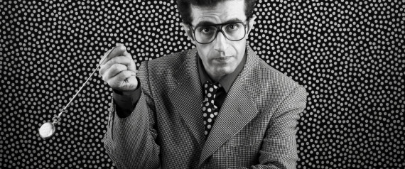
Hypnosis to help with sleeping problems

This article explains how hypnosis could help people with sleep problems.

Insomniacs and light sleepers alike might benefit from hypnosis to coax them into a deep sleep, suggests a new study published in the journal Sleep.
Researchers from the University of Zurich and Fribourg concluded that hypnosis can increase the quality of sleep, eliminating the need for medications the effects of which have long been questioned by the medical community.
"It opens up new, promising opportunities for improving the quality of sleep without drugs," says biopsychologist Björn Rasch, who headed the study at the Psychological Institute of the University of Zurich in conjunction with the Sleep and Learning project.
The study limited its participants to women, due to their increased susceptibility to hypnosis on average. A total of 70 healthy young women were divided into groups according to their susceptibility to hypnosis and invited to the sleep laboratory for a 90-minute midday nap.
The test group listened to 13 minutes of pre-recorded hypnosis before sleeping, and the control group listened to neutral dialogue over loudspeakers.
The most hypnotizable participants experienced an 80 per cent increase in slow wave sleep.
"The results may be of major importance for patients with sleep problems and for older adults," says psychologist and study leader Maren Cordi. "In contrast to many sleep-inducing drugs, hypnosis has no adverse side effects."
The findings indicate that even healthy individuals with a delayed circadian preference could further increase their health by taking steps to get to sleep earlier.
Deep sleep is referred to in the scientific community as slow wave sleep (SWS) and is measured by the actual speed of the brainwaves.
It is understood to be the most restorative of the sleep phases, for during SWS growth hormones are secreted, promoting cell repair and stimulating the body's immune system.
Read more here

Sunday, July 22, 2012

Epileptic encephalopathies of the Landau-Kleffner and continuous spike and waves during slow-wave sleep types: Genomic dissection makes the link with autism


Epileptic encephalopathies of the Landau-Kleffner


 and continuous spike and waves during slow-wave 


sleep types: 


Genomic dissection makes the link with autism


Summary


Purpose:  The continuous spike and waves during slow-wave sleep syndrome (CSWSS) and the Landau-Kleffner (LKS) syndrome are two rare epileptic encephalopathies sharing common clinical features including seizures and regression. Both CSWSS and LKS can be associated with the electroencephalography pattern of electrical status epilepticus during slow-wave sleep and are part of a clinical continuum that at its benign end also includes rolandic epilepsy (RE) with centrotemporal spikes. The CSWSS and LKS patients can also have behavioral manifestations that overlap the spectrum of autism disorders (ASD). An impairment of brain development and/or maturation with complex interplay between genetic predisposition and nongenetic factors has been suspected. A role for autoimmunity has been proposed but the pathophysiology of CSWSS and of LKS remains uncharacterized.
Methods:  In recent years, the participation of rare genomic alterations in the susceptibility to epileptic and autistic disorders has been demonstrated. The involvement of copy number variations (CNVs) in 61 CSWSS and LKS patients was questioned using comparative genomic hybridization assays coupled with validation by quantitative polymerase chain reaction (PCR).
Key Findings:  Whereas the patients showed highly heterogeneous in genomic architecture, several potentially pathogenic alterations were detected. A large number of these corresponded to genomic regions or genes (ATP13A4CDH9CDH13CNTNAP2CTNNA3,DIAPH3GRIN2AMDGA2SHANK3) that have been either associated with ASD for most of them, or involved in speech or language impairment, or in RE. Particularly, CNVs encoding cell adhesion proteins (cadherins, protocadherins, contactins, catenins) were detected with high frequency (≈20% of the patients) and significant enrichment (cell adhesion: p = 0.027; cell adhesion molecule binding: p = 9.27 × 10−7).
Significance:  Overall our data bring the first insights into the possible molecular pathophysiology of CSWSS and LKS. The overrepresentation of cell adhesion genes and the strong overlap with the genetic, genomic and molecular ASD networks, provide an exciting and unifying view on the clinical links among CSWSS, LKS, and ASD.