Showing posts with label absence seizure. Show all posts

Tuesday, June 10, 2014

Neural transplant reduces seizures in mice

A neural transplant reduces a specific type of
epileptic seizures in mice.

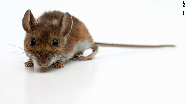
New research from North Carolina State University pinpoints the areas of the cerebral cortex that are affected in mice with absence epilepsy and shows that transplanting embryonic neural cells into these areas can alleviate symptoms of the disease by reducing seizure activity. The work may help identify the areas of the human brain affected in absence epilepsy and lead to new therapies for sufferers.
Absence epilepsy primarily affects children. These seizures differ from "clonic-tonic" seizures in that they don't cause muscle spasms; rather, patients "zone out" or stare into space for a period of time, with no memory of the episode afterward. Around one-third of patients with absence epilepsy fail to respond to medication, demonstrating the complexity of the disease.
NC State neurobiology professor Troy Ghashghaei and colleagues looked at a genetic mouse model for absence epilepsy to determine what was happening in their brains during these seizures. They found that the seizures were accompanied by hyperactivity in the areas of the brain associated with vision and touch -- areas referred to as primary visual and primary somatosensory cortices in the occipital and parietal lobes, respectively.
"There are neurons that excite brain activity, and neurons that inhibit activity," Ghashghaei says. "The inhibitory neurons work by secreting an inhibitory neurotransmitter called gamma-aminobutyric acid, or GABA. The 'GABAergic' interneurons were recently shown by others to be defective in the mice with absence seizures, and we surmised that these malfunctioning neurons might be part of the problem, especially in the visual and somatosensory cortical areas."
Ghashghaei's team took embryonic neural stem cells from a part of the developing brain that generates GABAergic interneurons for the cerebral cortex. They harvested these cells from normal mouse embryos and transplanted them into the occipital cortex of the genetic mice with absence seizures. Absence seizure activity in treated animals decreased dramatically, and the mice gained more weight and survived longer than untreated mice.
"This is a profound and remarkably effective first result, and adds to the recent body of evidence that these transplantation treatments can work in mouse models of epilepsy. But we still don't understand the mechanisms behind what the normal inhibitory cells are doing in areas of the visual cortex of absence epileptic mice," Ghashghaei says. "We know that you can get positive results even when a small number of transplanted neurons actually integrate into the cortex of affected mice, which is very interesting. But we don't know how the transplanted cells are connecting with other cells in the cortex and how they alleviate the absence seizures in the mouse model we employed.
"Our next steps will be to explore these questions. In addition, we are very interested in methods being devised by multiple labs around the world to 'reprogram' cells from transplantation patients to generate normal GABAergic and other types of neurons. Once established, this would eliminate the need for embryonic stem cells for this type of treatment. The ultimate goal is to develop new therapies for humans suffering from various forms of epilepsies, especially those for whom drugs do not work."
The research appears online in Cerebral Cortex.
Read more here

Monday, October 28, 2013

Prevalence of EEG paroxysmal activity in a population of children with obstructive sleep apnea syndrome.


SleepSleepSleep 2009; 32(4)

Prevalence of EEG paroxysmal activity in a population of children with obstructive sleep apnea syndrome.

Silvia Miano, Maria Chiara Paolino, Rosa Adrados, Marilisa Montesano, Salvatore Barberi, Maria Pia Villa
View via PMCView Original ArticleLikeCommentShare

STUDY OBJECTIVES:

Sleep breathing disorders may trigger paroxysmal events during sleep such as parasomnias and may exacerbate preexisting seizures. We verified the hypothesis that the amount of EEG paroxysmal activity (PA) may be high in children with obstructive sleep apnea syndrome (OSAS).

DESIGN:

Prospective study.

SETTINGS:

Sleep unit of an academic center.

PARTICIPANTS:

Polysomnographic studies were performed in a population of children recruited prospectively, for suspected OSAS, from January to December 2007, with no previous history of epileptic seizures or any other medical conditions. All sleep studies included > or = 8 EEG channels, including centrotemporal leads. We collected data about clinical and respiratory parameters of children with OSAS and with primary snoring, then we performed sleep microstructure analysis in 2 OSAS subgroups, matched for age and sex, with and without paroxysmal activity.

MEASUREMENTS AND RESULTS:

We found 40 children who met the criteria for primary snoring, none of them showed PA, while 127 children met the criteria for OSAS and 18 of them (14.2%) showed PA. Children with PA were older, had a predominance of boys, a longer duration of OSAS, and a lower percentage of adenotonsillar hypertrophy than children without PA. Moreover, PA occurred over the centrotemporal regions in 9 cases, over temporal-occipital regions in 5, and over frontocentral regions in 4. Children with PA showed a lower percentage of REM sleep, a lower CAP rate and lower A1 index during slow wave sleep, and lower total A2 and arousal index than children without EEG abnormalities.

CONCLUSIONS:

We found a higher percentage of paroxysmal activity in children with OSAS, compared to children with primary snoring, who did not exhibit EEG abnormalities. The children with paroxysmal activity have peculiar clinical and sleep microstructure characteristics that may have implications in the neurocognitive outcome of OSAS.

Sunday, March 17, 2013

What are types of seizures? How do you recognize seizures?

This article offers an overview of the different types of epileptic seizures and how to recognize them. JR


Epilepsy, also commonly known as a seizure disorder, is a medical condition that affects mental and physical functions.
Epilepsy causes clusters of nerves in the brain to work abnormally, which causes recurrent seizures. A person is often diagnosed with epilepsy if he or he has at least two unprovoked seizures.
According to the Centers for Disease Control and Prevention, 2.2 million Americans have epilepsy. Epilepsy can become more common as people age. However, lifetime risk of developing epilepsy is 4 percent. The goal of treatment is to render patients seizure-free with antiepileptic medications.
At times, patients can still experience seizures despite being on medications. Epilepsy surgery and electronic stimulation (via vagal nerve stimulator) are safe alternatives for intractable epilepsy.
There are many different causes of epilepsy. Epilepsy can be genetic, caused by a prior head trauma, and can be seen in individuals with history of meningitis, encephalitis, brain tumor or stroke.
Types of epileptic seizures
General seizure: The entire brain is involved in this type of seizure. The person may make a noise and stiffen for several seconds with rhythmic movements of the arms and legs. The eyes usually remain open during the seizure. After the seizure is over, the person may act confused. This seizure also is known as a grand mal seizure.
Partial seizure: Only part of the brain is involved in this type of seizure, meaning only part of the body is affected. For example, if the part of the brain that controls hand movement is affected, the person’s hand may jerk. Symptoms of partial seizures can include small repetitive movements. After having a partial seizure, the person may act confused.
Absence seizure: Absence seizures are most common in children. Those affected by this type of seizure often stare blankly and can blink repetitively. These seizures only last a few moments, and some people may have many of these in one day.
When diagnosing epilepsy, physicians often use an electroencephalogram, or EEG. This test records the electrical activity in the brain. People with epilepsy will often have changes in brain wave patterns, even when not having a seizure. Diagnosis is aided by having a CT, MRI or PET scan. The scans can find abnormalities in the brain that might be causing seizures.
Anyone experiencing a seizure for the first time should see a doctor immediately.
Read more here