Showing posts with label brain connections. Show all posts

Sunday, May 10, 2015

Study: Premature birth linked to altered brain connectivity and developmental disorders

According to a recent study, premature birth is linked to altered brain connectivity.- JR

Premature birth can alter the connectivity between key areas of the brain, according to a new study led by King's College London. The findings should help researchers to better understand why premature birth is linked to a greater risk of neurodevelopmental problems, including autistic spectrum disorders and attention deficit disorders.

The NIHR-funded study, published in the journal PNAS, used functional magnetic resonance imaging (fMRI) to look at specific connections in the brains of 66 infants, 47 of whom were born before 33 weeks and were therefore at high risk of neurological impairment, and 19 born at term. The brain connections investigated were between the thalamus and the cortex, connections which develop rapidly during the period a preterm infant is cared for on a neonatal unit.

Researchers found that those born in the normal window of birth (37-42 weeks) showed a remarkably similar structure to adults in these brain regions, strengthening existing evidence that the brain's network of connections is quite mature at the time of birth.

However, infants born prematurely (before 33 weeks gestation) were found to have less connectivity between areas of the thalamus and particular areas of the brain's cortex known to support higher cognitive functions, but greater connectivity between the thalamus and an area of primary sensory cortex which is involved in processing signals from the face, lips, jaw, tongue, and throat.
The greater the extent of prematurity, the more marked were the differences in the pattern of brain connectivity.

The authors suggest that the stronger connections involving face and lips in babies born preterm may reflect their early exposure to breastfeeding and bottlefeeding, while the reduced connectivity in other brain regions may be linked to the higher incidence of difficulties seen in later childhood.
Dr Hilary Toulmin, first author from the Centre for the Developing Brain at King's College London, said: 'The next stage of our work will be to understand how these findings relate to the learning, concentration and social difficulties which many of these children experience as they grow older.'
Professor David Edwards, senior author from the Centre for the Developing Brain at King's College London, said: 'The ability of modern science to image the connections in the brain would have been inconceivable just a few years ago, but we are now able to observe brain development in babies as they grow, and this is likely to produce remarkable benefits for medicine.'
Read more here

Tuesday, December 03, 2013

Connections within young children's brains strengthen while they sleep

A study shows that as children under five years old sleep, the connections between the left and right halves of their brain strengthen showing the importance of sleep in young children.

As young children sleep, the connections between the right and left sides of their brains strengthen, according to a small new study.
Researchers measured the brain activity of eight children while they slept at ages 2, 3 and 5 years. They found that connections in the brain generally became stronger during sleep as the children aged.
The strength of the connections between the left and right sides of the brain increase as much as 20 percent over a night's sleep, according to the study, which was published online Nov. 12 in the journal Brain Sciences.
"There are strong indications that sleep and brain maturation are closely related, but at this time, it is not known how sleep leads to changes in brain structure," study leader Salome Kurth, a postdoctoral researcher at the University of Colorado, Boulder, said in a university news release.
How sleep disruption during childhood may affect brain development and behavior will be examined in future studies.
"I believe inadequate sleep in childhood may affect the maturation of the brain related to the emergence of developmental or mood disorders," Kurth said.
It was already known that the brain changes drastically during early childhood. New connections are formed, others disappear and a fatty layer called "myelin" forms around nerve fibers in the brain. The growth of myelin strengthens the connections by speeding up the transfer of information, according to background information included in the news release.
Maturation of nerve fibers improves children's skills in areas such as language, attention and impulse control. But it hasn't been clear what role sleep plays in the development of such brain connections.
Read more here

Thursday, August 22, 2013

Brain differences in those with Autism: Think Local Not Global

Fig. 4.A new study shows that a person with autism's brain is wired differently than those without autism, and this finding may help explain some of the typical symptoms of autism. 

Increased connectivity may favor local connections over global. - JR

The way the gray matter in the brain is wired appears to be different in people with autism, new research shows.
Specifically, those with the disorder are more likely to have enhanced connections in the brain that are associated with common autism symptoms, such as narrow interests and repetitive behaviors, the scientists reported.
"Our study [and others] reliably and repeatedly demonstrate that the brain in autism is built and functions differently, which explains a variety of autistic symptoms," said study author Christine Ecker, a lecturer in neuroimaging at King's College London.
"From neuropsychology, we also know that people with autism often have a preference for processing [fine details] over global features (the ability to integrate piecemeal information into a coherent whole)," she said. "Our findings may therefore represent a neuroanatomical correlate of these behaviors, although a direct causal link remains to be established."
Results of the study were published online July 22 in the Proceedings of the National Academy of Sciences.
Autism spectrum disorders are a group of neurodevelopmental disorders, common symptoms of which include impaired social communication, social reciprocity and repetitive behaviors, according to background information included in the study. Experts believe that differences in the brain account for these behaviors, but the exact changes that might occur in someone with autism aren't yet clear, according to the study.
The current study looked at 34 adult males with autism and 34 males without autism to serve as the control group. All of the study volunteers underwent MRI.
The researchers found that there were significant differences in the length of the connections between regions of the brain when they compared people with autism to those without. The minimum length of these connections in the cortical gray matter was dubbed "wiring costs" by the researchers. These wiring costs were significantly reduced in people with autism, meaning the lengths of their connections were shorter.
"These differences are predominantly observed in brain regions that we know are anatomically different in autism, and that are underlying autistic symptoms and traits," Ecker said. "We think that such differences in neuronal wiring may lead to locally over-connected networks in the brains of [patients with autism spectrum disorders] that could explain some autistic symptoms, such as repetitive behaviors."
One expert said the findings help define the biological basis of autism more clearly.
"We've known that there is a history of wiring differences that appear in individuals with autism. What's novel is that they used structural imaging to assess whether neurons are connected in the same way structurally," said Daniel Smith, senior director of discovery neuroscience at Autism Speaks.
"Gray matter connections are referred to as microcircuits, and they're everywhere," Smith said. "They're very important in the cerebral cortex, and the highest levels of thinking occur in the cerebral cortical regions of the brain."
"This study is another component in building our knowledge base," he added. "It's a step toward better understanding of what's happening in the brain and, ultimately, that will help lead to new treatments. But this study won't lead to an immediate impact on treatment."
Dr. Andrew Adesman, chief of developmental and behavioral pediatrics at the Steven and Alexandra Cohen Children's Medical Center in New Hyde Park, N.Y., agreed that this study won't lead to any immediate changes in the field.
"Unfortunately, despite the many advances in our ability to study the brain and identify a range of structural and functional differences associated with autism, we are still left with more questions than answers," Adesman said.
"[But] studies such as this bring us one step closer to understanding the neurobiological underpinnings of the mysterious and enigmatic condition known as autism spectrum disorder," he said.
Ecker said she hopes to conduct a study that follows young people as their brains are developing to see how the connections in the brain behave over time in people with autism.
Read more here

Full article

Sunday, March 03, 2013

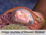
Connections in the Developing Brains of 3rd Trimester Babies

Scientists found the order and strength of brain connections in developing fetuses. This information may be used to understand how brain connections can go wrong leading to disorders such as dyslexia, ADHD, and autism.

Using real-time images of brain connections developing in late-stage fetuses, scientists say they've been able for the first time to compare the order and strength of these connections.
The research, though very preliminary, might one day lead the way to more effective therapies for brain disorders such as dyslexia, attention-deficit/hyperactivity disorder (ADHD) and autism, the researchers said.
Scientists from the U.S. National Institute of Child Health and Human Development and Wayne State University School of Medicine looked at 25 fetal brains from a group of pregnant women between 24 and 38 weeks of gestation. The researchers used an imaging technique called functional MRI (fMRI) to visualize "communication" between various regions of the brain.
The scientists learned that connections between the right and left sides of the brains got stronger as fetuses grew older. They said they also learned that shorter distances between matching areas produced stronger signals than longer spans between corresponding areas on the brain's outer edges.
"What we're seeing is a picture of emerging connectivity . . . that the right and left side are kind of building a bridge to each other," said study author Moriah Thomason, an assistant professor of pediatrics at Wayne State, in Detroit. "It's evidence for the fact that already in fetal life, this anatomy is being constructed in a way that we would expect. But for the first time, we can show the development of these networks."
The findings are published in the Feb. 20 issue of the journal Science Translational Medicine.
While scientists had previously used fMRI scans on fetuses, this new research is the first to compare a group of fetuses and identify features of development, Thomason said. Nearly 90 percent of the pregnant mothers participating in the study were black, while the rest were white or multiracial. They later delivered 17 boys and eight girls.
MRI scans don't emit radiation, thus minimizing risk to the fetuses during the procedure. The technique showed significant connections between half of the dozens of brain areas tested.
The findings might provide groundwork for understanding how and when brain development may go awry during gestation, Thomason said. This may possibly lead to better understanding of conditions such as ADHD, dyslexia and autism, which are thought to arise from disrupted brain networks, she said.
"If we know what gets in the way of those [normal] processes, we have a better shot at treating those disorders," Thomason said. "It's not just about early identification. An additional valuable outcome is, when you can see what normal looks like and see what disruption looks like, you have the opportunity to pick out patterns that tell you about the origin of that disease," she explained.
"The brain can be a tattletale to what is going on in those diseases," Thomason added, "and that can help us develop novel treatments."
Read more here